Differential Diagnosis of Knee Pain Commonly Encountered in Clinical Practice
- Affiliations
-
- 1Joint Reconstruction Center, Seoul National University Bundang Hospital, Seoul National University College of Medicine, Seongnam, Korea. drchuc@chol.com
- 2Department of Orthopaedic Surgery, Seoul National University Bundang Hospital, Seoul National University College of Medicine, Seongnam, Korea.
- KMID: 2202166
- DOI: http://doi.org/10.4078/jkra.2007.14.3.185
Abstract
- The knee is a complex structure and far from being the simple hinge joint of popular belief. Knee pain would not be properly understood unless one is familiar with the anatomy and understands the role of the various structures. For patient consultation or for selecting a treatment option, it is crucial to find the central cause of symptoms and functional disabilities of the patient based on a detailed history, a focused examination and, when indicated, the selective use of appropriate imaging and laboratory studies. The history taking should be comprehensive and include the demographic characteristics, past medical history, comprehending the associated trauma, pain characteristics and quality of life. Basic physical examination should include inspection of walking pattern and the knee, evaluation of joint effusion, range of motion and the location of tenderness, and precise assessment of joint stability. Although many advanced diagnostic tools are available, plain radiographs are frequently utilized as a primary tool to evaluate conditions of the knee joint for practical and economic reasons. A weight-bearing anteroposterior radiographs should be taken for appropriate evaluation of the condition of the tibiofemoral joint, and weight-bearing radiographs in semi-flexed position is valuable to evaluate the joint space more precisely. To evaluate the patellofemoral joint, axial and lateral views should be included in the routine radiographs. In practice, physicians need to be cautious not to easily reach the conclusion that the symptoms of the knee joint can be attributed to only a few clinical findings in consulting the patients with knee pain.
Keyword
MeSH Terms
Figure
-

Fig. 1. Standing anteroposterior radiograph in extension (A) and standing 45o flexion posteroanterior radiograph (B) of a 69-year-old female who suffered from left knee pain. The radiograph in A indicates mild joint space narrowing (arrow), while the radiograph in B indicates almost obliteration of the joint space (arrow).

Fig. 2. Standing whole-length lower leg anteroposterior radiograph for evaluation of the limb alignment. In the radiograph, the lines connecting the center of the femoral head and the talus passed through the medial side of both knee joints, indicating significant varus alignment of the lower leg.
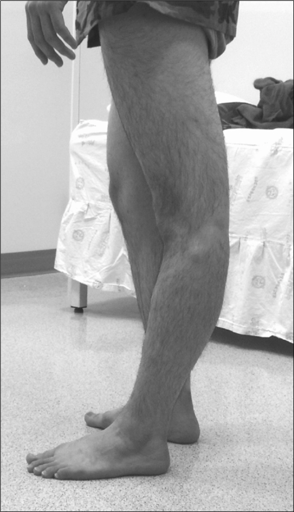
Fig. 3. A 21-year old man with posterlateral corner injury of the left knee. The photograph shows hyperextension of the left knee.

Fig. 4. Standing anteroposterior radiographs of a 62-year-old female who suffered from right knee pain. (A) A radiograph taken at her first visit to our institution shows only minimal osteoarthritic change of the knee joint. (B) A radiograph taken 8 months after her first visit shows a large osteonecrotic lesion in the medial femoral condyle (white arrow). (C) An intraoperative photograph shows osteonecrotic lesion involved the medial femoral condyle (black arrow).
Reference
-
1). Insall J., Scott W. Surgery of knee. 3rd ed.p. 86–98. 145-192, 615-714. Philadelphia, Churchill Livingstone;2001.2). Chapman M. Chapman's orthopaedic surgery. 3rd ed.p. 2247–2268. 2299-2310, 2393-2434. Philadelphia, Lippincott Williams & Wilkins;2001.3). Rosenberg TD., Paulos LE., Parker RD., Coward DB., Scott SM. The forty-five-degree posteroanterior flexion weight-bearing radiograph of the knee. J Bone Joint Surg Am. 1988. 70:1479–83.
Article4). Leach RE., Gregg T., Siber FJ. Weight-bearing radiography in osteoarthritis of the knee. Radiology. 1970. 97:265–8.
Article5). Inoue S., Nagamine R., Miura H., Urabe K., Matsuda S., Sakaki K, et al. Anteroposterior weight-bearing radiography of the knee with both knees in semiflexion, using new equipment. J ᄋrthop Sci. 2001. 6:475–80.
Article6). Fulkerson J. Disoders of the Patellofemoral Joint. 4th ed.p. 76–106. 185-210. Philadelphia, Lippincott Williams and Wilkins;2004.7). Woo SL., Inoue M., McGurk-Burleson E., Gomez MA. Treatment of the medial collateral ligament injury. II: Structure and function of canine knees in response to differing treatment regimens. Am J Sports Med. 1987. 15:22–9.8). Kanamori A., Woo SL., Ma CB., Zeminski J., Rudy TW., Li G, et al. The forces in the anterior cruciate ligament and knee kinematics during a simulated pivot shift test: a human cadaveric study using robotic technology. Arthroscopy. 2000. 16:633–9.
Article9). Lerat JL., Moyen BL., Cladiere F., Besse JL., Abidi H. Knee instability after injury to the anterior cruciate ligament. Quantification of the Lachman test. J Bone Joint Surg Br. 2000. 82:42–7.10). Micheo W., Frontera WR., Amy E., Jordan G. Rehabilitation of the patient with an anterior cruciate ligament injury: a brief review. Bol Asoc Med P R. 1995. 87:29–36.11). Muaidi QI., Nicholson LL., Refshauge KM., Herbert RD., Maher CG. Prognosis of conservatively managed anterior cruciate ligament injury: a systematic review. Sports Med. 2007. 37:703–16.12). Fu FH., Bennett CH., Lattermann C., Ma CB. Current trends in anterior cruciate ligament reconstruction. Part 1: Biology and biomechanics of reconstruction. Am J Sports Med. 1999. 27:821–30.13). Fu FH., Bennett CH., Ma CB., Menetrey J., Lattermann C. Current trends in anterior cruciate ligament reconstruction. Part II. Operative procedures and clinical correlations. Am J Sports Med. 2000. 28:124–30.14). Canale S. Campbell's operative orthopaedics. 10th ed.p. 2253–2301. Philadelphia, Mosby-year Book Inc;2003.15). Shelbourne KD., Gray T. Natural history of acute posterior cruciate ligament tears. J Knee Surg. 2002. 15:103–7.16). Torg JS., Barton TM., Pavlov H., Stine R. Natural history of the posterior cruciate ligament-deficient knee. Clin ᄋrthop Relat Res. 1989. 246:208–16.
Article17). Dennis MG., Fox JA., Alford JW., Hayden JK., Bach BR Jr. Posterior cruciate ligament reconstruction: current trends. J Knee Surg. 2004. 17:133–9.18). Zhao J., Huangfu X. Arthroscopic singlebundle posterior cruciate ligament reconstruction: Retrospective review of 4-versus 7-strand hamstring tendon graft. Knee. 2007. 14:301–5.19). Krudwig WK., Witzel U., Ullrich K. Posterolateral aspect and stability of the knee joint. II. Posterolateral instability and effect of isolated and combined posterolateral reconstruction on knee stability: a biomechanical study. Knee Surg Sports Traumatol Arthrosc. 2002. 10:91–5.
Article20). LaPrade RF., Ly TV., Wentorf FA., Engebretsen L. The posterolateral attachments of the knee: a qualitative and quantitative morphologic analysis of the fibular collateral ligament, popliteus tendon, popliteofibular ligament, and lateral gastrocnemius tendon. Am J Sports Med. 2003. 31:854–60.21). DiStefano VJ. Function, post-traumatic sequelae and current concepts of management of knee meniscus injuries: a review article. Clin ᄋrthop Relat Res. 1980. 151:143–6.22). Fox MG. MR imaging of the meniscus: review, current trends, and clinical implications. Magn Reson Imaging Clin N Am. 2007. 15:103–23.
Article23). Sgaglione NA. Meniscus repair update: current concepts and new techniques. Orthopedics. 2005. 28:280–6.
Article24). Clanton TO., DeLee JC. Osteochondritis dissecans. History, pathophysiology and current treatment concepts. Clin ᄋrthop Relat Res. 1982. 167:50–64.
Article25). Cahill BR. Current concepts review. Osteochondritis dissecans. J Bone Joint Surg Am. 1997. 79:471–2.26). Kocher MS., Tucker R., Ganley TJ., Flynn JM. Management of osteochondritis dissecans of the knee: current concepts review. Am J Sports Med. 2006. 34:1181–91.27). Mochizuki T., Akita K., Muneta T., Sato T. Pes anserinus: layered supportive structure on the medial side of the knee. Clin Anat. 2004. 17:50–4.
Article28). Uson J., Aguado P., Bernad M., Mayordomo L., Naredo E., Balsa A, et al. Pes anserinus tendino-bursitis: what are we talking about? Scand J Rheumatol. 2000. 29:184–6.29). Kim JY., Finger DR. Spontaneous osteonecrosis of the knee. J Rheumatol. 2006. 33:1416.30). Narvaez JA., Narvaez J., De Lama E., Sanchez A. Spontaneous osteonecrosis of the knee associated with tibial plateau and femoral condyle insufficiency stress fracture. Eur Radiol. 2003. 13:1843–8.31). Satku K., Kumar VP., Chong SM., Thambyah A. The natural history of spontaneous osteonecrosis of the medial tibial plateau. J Bone Joint Surg Br. 2003. 85:983–8.
Article32). Yamamoto T., Bullough PG. Spontaneous osteonecrosis of the knee: the result of subchondral insufficiency fracture. J Bone Joint Surg Am. 2000. 82:858–66.
Article33). Mont MA., Rifai A., Baumgarten KM., Sheldon M., Hungerford DS. Total knee arthroplasty for osteonecrosis. J Bone Joint Surg Am. 2002. 84-A:599–603.
Article34). Myers TG., Cui Q., Kuskowski M., Mihalko WM., Saleh KJ. Outcomes of total and unicompartmental knee arthroplasty for secondary and spontaneous osteonecrosis of the knee. J Bone Joint Surg Am. 2006. 88(Suppl 3):76–82.
Article35). Dye SF. The knee as a biologic transmission with an envelope of function: a theory. Clin ᄋrthop Relat Res. 1996. 325:10–8.36). Dye SF. The pathophysiology of patellofemoral pain: a tissue homeostasis perspective. Clin ᄋrthop Relat Res. 2005. 436:100–10.37). E LL., Padron M. Anterior knee pain. Eur J Radiol. 2007. 62:27–43.38). Amis AA. Current concepts on anatomy and biomechanics of patellar stability. Sports Med Arthrosc. 2007. 15:48–56.
Article39). Mulford JS., Wakeley CJ., Eldridge JD. Assessment and management of chronic patellofemoral instability. J Bone Joint Surg Br. 2007. 89:709–16.
Article40). Sanchis-Alfonso V., Rosello-Sastre E., MartinezSanjuan V. Pathogenesis of anterior knee pain syndrome and functional patellofemoral instability in the active young. Am J Knee Surg. 1999. 12:29–40.41). Grelsamer RP. Patellar nomenclature: the Tower of Babel revisited. Clin ᄋrthop Relat Res. 2005. 436:60–5.42). Arendt EA., Fithian DC., Cohen E. Current concepts of lateral patella dislocation. Clin Sports Med. 2002. 21:499–519.
Article43). Roux C. Recurrent dislocation of the patella: operative treatment. 1888. Clin ᄋrthop Relat Res. 2006. 452:17–20.44). Vainionpaa S., Laasonen E., Silvennoinen T., Vasenius J., Rokkanen P. Acute dislocation of the patella. A prospective review of operative treatment. J Bone Joint Surg Br. 1990. 72:366–9.
Article45). Greis PE., Holmstrom MC., Lahav A. Surgical treatment options for patella tendon rupture, Part I: Acute. Orthopedics. 2005. 28:672–9.
Article46). Moonot P., Fazal MA. Traumatic patella tendon rupture: early mobilisation following surgical repair. Injury. 2005. 36:1385.
Article47). Bloom ᄋJ., Mackler L., Barbee J. Clinical inquiries. What is the best treatment for ᄋsgood-Schlatter disease? J Fam Pract. 2004. 53:153–6.48). Soren A. Treatment of ᄋsgood-Schlatter disease. Am J ᄋrthop Surg. 1968. 10:70–1.49). Jackson AM. Anterior knee pain. J Bone Joint Surg Br. 2001. 83:937–48.
Article50). Medlar RC., Lyne ED. Sinding-Larsen-Johansson disease. Its etiology and natural history. J Bone Joint Surg Am. 1978. 60:1113–6.
Article51). Matheson Gᄋ. Long-term prognosis for jumper's knee. Clin J Sport Med. 2003. 13:196.52). Pierets K., Verdonk R., De Muynck M., Lagast J. Jumper's knee: postoperative assessment. A retrospective clinical study. Knee Surg Sports Traumatol Arthrosc. 1999. 7:239–42.53). Schmid MR., Hodler J., Cathrein P., Duewell S., Jacob HA., Romero J. Is impingement the cause of jumper's knee? Dynamic and static magnetic resonance imaging of patellar tendinitis in an open-configuration system. Am J Sports Med. 2002. 30:388–95.54). Canizares GH., Selesnick FH. Bipartite patella fracture. Arthroscopy. 2003. 19:215–7.
Article55). Ireland ML., Chang JL. Acute fracture bipartite patella: case report and literature review. Med Sci Sports Exerc. 1995. 27:299–302.56). Kim SJ., Jeong JH., Cheon YM., Ryu SW. MPP test in the diagnosis of medial patellar plica syndrome. Arthroscopy. 2004. 20:1101–3.
Article57). Tindel NL., Nisonson B. The plica syndrome. ᄋrthop Clin North Am. 1992. 23:613–8.
Article58). Bentley G., Dowd G. Current concepts of etiology and treatment of chondromalacia patellae. Clin ᄋrthop Relat Res. 1984. 189:209–28.59). ᄋgilvie-Harris DJ., Jackson RW. The arthroscopic treatment of chondromalacia patellae. J Bone Joint Surg Br. 1984. 66:660–5.60). Thomee R., Augustsson J., Karlsson J. Patellofemoral pain syndrome: a review of current issues. Sports Med. 1999. 28:245–62.
- Full Text Links
-
- Actions
-
Cited
- CITED
-
- Close
- Share
-
- Similar articles
-
- Differential Diagnosis of Elbow Pain
- A Simple Working Classification Proposed for Orofacial Pain (OFP) Commonly Encountered in Dental Practice
- Up-To-Date Knowledge for Foot Disorders
- Rehabilitation of Sports-related Knee Injury
- Guidelines for the Management of Postoperative Pain after Total Knee Arthroplasty
