Primary Acinic Cell Carcinoma of the Lung: A Case Report
- Affiliations
-
- 1Department of Pathology, Samsung Medical Center, Sungkyunkwan University School of Medicine, Seoul, Korea. hanjho@skku.edu
- 2Department of Thoracic Surgery, Samsung Medical Center, Sungkyunkwan University School of Medicine, Seoul, Korea.
- 3Department of Radiology, Samsung Medical Center, Sungkyunkwan University School of Medicine, Seoul, Korea.
- KMID: 2200025
- DOI: http://doi.org/10.6058/jlc.2010.9.1.20
Abstract
- Primary acinic cell carcinoma (ACC) of the lung is very rare and this tumor is thought to arise from pluripotent cells of the submucosal glands of the tracheobronchial tree. We report here on a case of primary ACC of the lung in a 68-year-old man who had a solitary pulmonary nodule in the left lower lobe. The patient was symptomless and the lesion was found on a chest X-ray taken during a regular health checkup. The video assisted thoracoscopic surgery wedge resection revealed an ovoid yellow tan solid mass that was 1.8 cm at the largest diameter. Microscopically, the neoplastic cells grew in solid sheets of round cells with eccentric nuclei and abundant basophilic granular cytoplasm. There were no mitotic figures or areas of pleomorphic or anaplastic cells. Immunohistochemical staining for cytokeratin (AE1/AE3) was positive, but the staining for chromogranin A and CD56 was negative. Ultrastructural examination revealed polyhedral cells with many zymogen granules of varying electron density. The patient is well 4 months postoperatively.
MeSH Terms
Figure
-
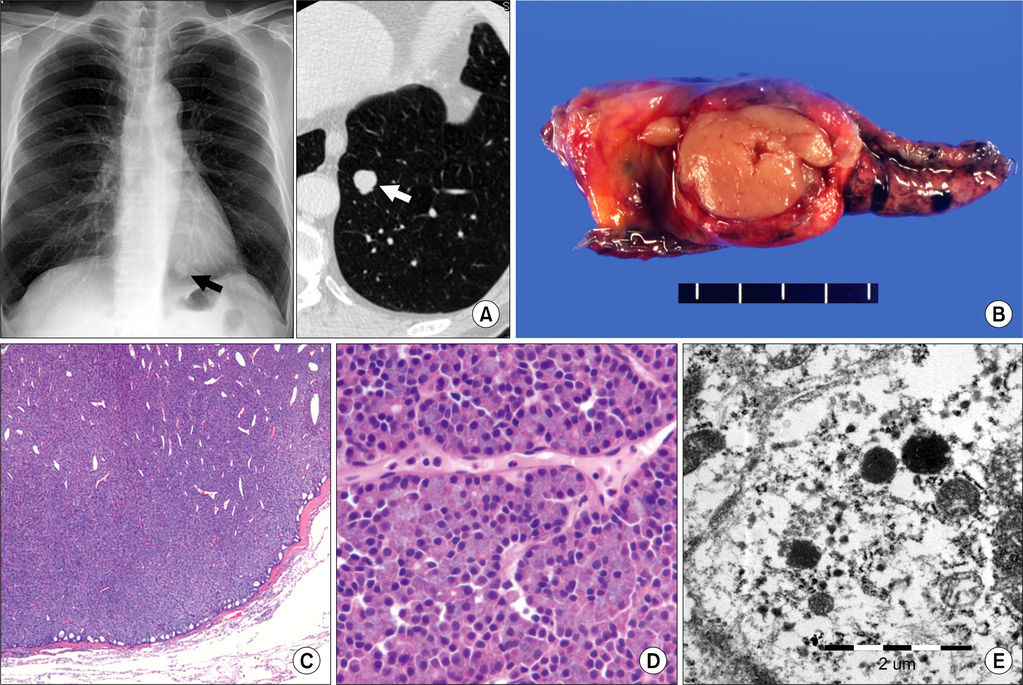
Fig. 1. (A) The chest PA X-ray and CT show an ovoid pulmonary nodule (1.3 cm, arrows) in the left lower lobe of lung. (B) On the VATS wedge resection, the cut surface of the lung shows a well demarcated round yellow tan solid mass without necrosis or hemorrhage. (C) The tumor has a fibrous pseudocapsule and it is composed of sheets of round or ovoid uniform cells with a peripheral microcystic pattern (H&E stain, ×50). (D) The tumor cells have eosinophilic or basophilic granular cytoplasm with round to oval nuclei (H&E stain, ×400). (E) On ultrastructural examination of the formalin-fixed, paraffin-embedded tissue, the cytoplasm of the tumor cells has many well-developed organelles, including many mitochondria, endoplasmic reticulum, ribosomes and glycogen granules. Several membrane bounded electron dense secretory granules are also identified.
Reference
-
References
1. Ukoha OO, Quartararo P, Carter D, Kashgarian M, Ponn RB. Acinic cell carcinoma of the lung with metastasis to lymph nodes. Chest. 1999; 115:591–595.
Article2. Fechner RE, Bentinck BR, Askew JB Jr. Acinic cell tumor of the lung: a histologic and ultrastructural study. Cancer. 1972; 29:501–508.3. Katz DR, Bubis JJ. Acinic cell tumor of the bronchus. Cancer. 1976; 38:830–832.
Article4. Heard BE, Dewar A, Firmin RK, Lennox SC. One very rare and one new tracheal tumour found by electron microscopy: glomus tumour and acinic cell tumour resembling carcinoid tumours by light microscopy. Thorax. 1982; 37:97–103.
Article5. Gharpure KJ, Deshpande RK, Vlshweshvara RN, Raghu CR, Bhargava MK. Acinic cell tumour of the bronchus (a case report). Indian J Cancer. 1985; 22:152–156.6. Moran CA, Suster S, Koss MN. Acinic cell carcinoma of the lung (“Fechner tumor”): a clinicopathologic, immunohistochemical, and ultrastructural study of five cases. Am J Surg Pathol. 1992; 16:1039–1050.7. Horowitz Z, Kronenberg J. Acinic cell carcinoma of the trachea. Auris Nasus Larynx. 1994; 21:193–195.
Article8. Ansari MA, Marchevsky A, Strick L, Mohsenifar Z. Upper airway obstruction secondary to acinic cell carcinoma of the trachea: use of Nd: YAG laser. Chest. 1996; 110:1120–1122.9. Lee HY, Mancer K, Koong HN. Primary acinic cell carcinoma of the lung with lymph node metastasis. Arch Pathol Lab Med. 2003; 127:e216–e219.
Article10. Sabaratnam RM, Anunathan R, Govender D. Acinic cell carcinoma: an unusual cause of bronchial obstruction in a child. Pediatr Dev Pathol. 2004; 7:521–526.
Article11. Tsukayama S, Omura K, Kanehira E, et al. Acinic cell carcinoma of the trachea: report of a case. Surg Today. 2004; 34:764–768.
Article12. Watanabe K, Ono N, Hoshi T, Hanzawa M, Ishida T. Fine-needle aspiration cytology of bronchial acinic cell carcinoma: a case report. Diagn Cytopathol. 2004; 30:359–361.
Article13. Chuah KL, Yap WM, Tan HW, Koong HN. Recurrence of pulmonary acinic cell carcinoma. Arch Pathol Lab Med. 2006; 130:932–933.
Article14. von Biberstein SE, Spiro JD, Mancoll W. Acinic cell carcinoma of the nasal cavity. Otolaryngol Head Neck Surg. 1999; 120:759–762.
Article15. Crissman JD, Rosenblatt A. Acinous cell carcinoma of the larynx. Arch Pathol Lab Med. 1978; 102:233–236.16. Kallis S, Stevens DJ. Acinous cell carcinoma of the larynx. J Laryngol Otol. 1989; 103:638–641.
Article17. Schmitt FC, Ribeiro CA, Alvarenga S, Lopes JM. Primary acinic cell-like carcinoma of the breast: a variant with good prognosis? Histopathology. 2000; 36:286–289.18. Minic AJ. Acinic cell carcinoma arising in a parotid lymph node. Int J Oral Maxillofac Surg. 1993; 22:289–291.19. Moran CA. Primary salivary gland-type tumors of the lung. Semin Diagn Pathol. 1995; 12:106–122.
- Full Text Links
-
- Actions
-
Cited
- CITED
-
- Close
- Share
-
- Similar articles
-
- Acinic Cell Carcinoma of The Parotid Gland: A Case Report
- Fine Needle Aspiration Cytology of Acinic Cell Carcinoma of the Parotid Gland: A Case Report
- Fine Needle Aspiration Cytology of Papillary-Cystic Variant of Acinic Cell Carcinoma of Salivary Gland: A Case Report
- Acinic Cell Carcinoma Arising from Unusual Location: 3 Case Reports
- Primary Acinic Cell Carcinoma of the Breast: A Case Report with an Immunohistochemical and Ultrastructural Studies
