J Korean Acad Prosthodont.
2009 Apr;47(2):206-214. 10.4047/jkap.2009.47.2.206.
Photoelastic stress analysis of the mandibular unilateral free-end removable partial dentures according to the design
- Affiliations
-
- 1Department of Prosthodontics, College of Dentistry, Chosun University, Korea. proscwpark@naver.com
- KMID: 2195618
- DOI: http://doi.org/10.4047/jkap.2009.47.2.206
Abstract
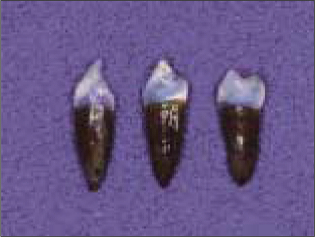
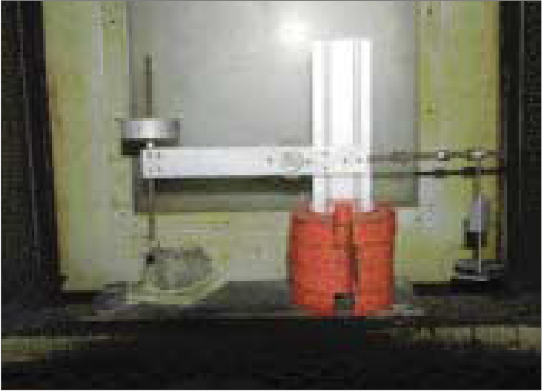
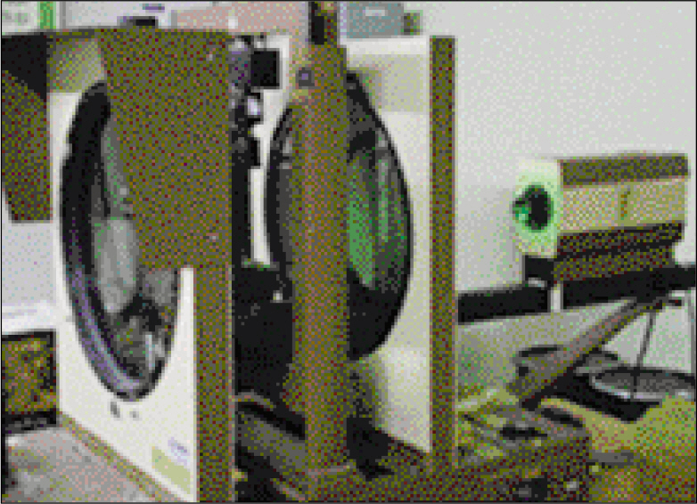
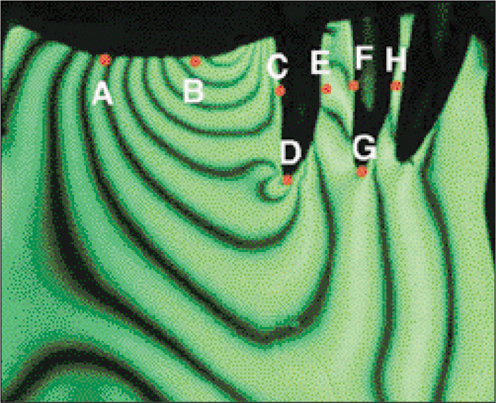
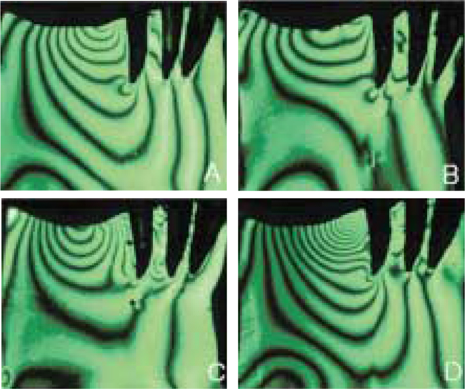
- STATEMENT OF PROBLEM: There are common clinical cases in which the mandibular first and second molars are missing unilaterally. PURPOSE: This study was designed to compare and evaluate the magnitude and distribution of stress produced by four kinds of mandibular unilateral free-end removable partial dentures that could be applied clinically in Kennedy class II cases. MATERIAL AND METHODS: Four unilateral free-end removable partial dentures using clasp, Konus crown, resilient attachment, and flexible resin were fabricated on the photoelastic models of the Kennedy class II cases. The vertical load of 6kg was applied on the central fossa of the first molar of every removable partial denture in the stress freezing furnace and the photoelastic models were frozen according to the stress freezing cycle. After these models were sliced mesio-distally to a thickness of 6mm, the photoelastic isochromatic white and black lines of the sliced specimens were examined with the transparent photoelastic experiment device and photographs were taken with a digital camera. The fringe order numbers at eight measuring points in the photograph were measured with the naked eye. RESULTS: The maximum fringe order number of each sliced specimen and the fringe order number at the residual ridge just below the loading point were in the decreasing order of the unilateral removable partial dentures using flexible resin followed by clasp, resilient attachment, and Konus crown. The fringe order number at the root apex of the second premolar was in the decreasing order of the unilateral removable partial dentures using clasp followed by flexible resin, Konus crown, and resilient attachment. CONCLUSION: The removable partial denture using Konus crown showed the most equalized stress distribution to the supporting alveolar bone of abutment teeth and residual ridge under the vertical loads. The removable partial denture using flexible resin can be applied to the case that has a better state of residual ridge than abutment teeth.
Keyword
Figure
Reference
-
1.Berg T., Caputo AA. Maxillary distal-extension removable partial denture abutments with reduced periodontal support. J Prosthet Dent. 1993. 70:245–50.
Article2.Park I., Eto M., Wakabayashi N., Hideshima M., Ohyama T. Dynamic retentive force of a mandibular unilateral removable partial denture framework with a back-action clasp. J Med Dent Sci. 2001. 48:105–11.3.Jin X., Sato M., Nishiyama A., Ohyama T. Influence of loading positions of mandibular unilateral distal extension removable partial dentures on movements of abutment tooth and denture base. J Med Dent Sci. 2004. 51:155–63.4.Vang MS. Case reports on the removable partial dentures with Konus telescope. J Korean Acad Prosthodont. 1997. 35:67–77.5.Kay KS., Shin HD., Song HN. Clinical cases & treatment conception for the distal extension removable partial denture using the attachment. Oral Biology Research. 2005. 29:113–34.6.Park CW., Hwang YP., Kay KS. Prosthetic restoration of partially edentulous patients using the Valplast® flexible partial denture system. Oral Biology Research. 2006. 30:55–73.7.Monteith BD. Management of loading forces on mandibular distal-extension prostheses. Part II: Classification for matching modalities to clinical situations. J Prosthet Dent. 1984. 52:832–6.
Article8.Browning JD., Meadors LW., Eick JD. Movement of three removable partial denture clasp assemblies under occlusal loading. J Prosthet Dent. 1986. 55:69–74.
Article9.Frechette AR. The influence of partial denture design on distribution of force to abutment teeth. 1956. J Prosthet Dent. 2001. 85:527–39.10.Kydd WL., Daly CH. The biologic and mechanical effects of stress on oral mucosa. J Prosthet Dent. 1982. 47:317–29.
Article11.Chou TM., Caputo AA., Moore DJ., Xiao B. Photoelastic analysis and comparison of force-transmission characteristics of intracoronal attachments with clasp distal-extension removable partial dentures. J Prosthet Dent. 1989. 62:313–9.
Article12.Reitz PV., Caputo AA. A photoelastic study of stress distribution by a mandibular split major connector. J Prosthet Dent. 1985. 54:220–5.
Article13.Langer A. Telescope retainers for removable partial dentures. J Prosthet Dent. 1981. 45:37–43.
Article14.Igarashi Y., Ogata A., Kuroiwa A., Wang CH. Stress distribution and abutment tooth mobility of distal-extension removable partial dentures with different retainers: an in vivo study. J Oral Rehabil. 1999. 26:111–6.15.Kratochvil FJ., Thompson WD., Caputo AA. Photoelastic analysis of stress patterns on teeth and bone with attachment retainers for removable partial dentures. J Prosthet Dent. 1981. 46:21–8.
Article16.Saito M., Miura Y., Notani K., Kawasaki T. Stress distribution of abutments and base displacement with precision attachmentand telescopic crown-retained removable partial dentures. J Oral Rehabil. 2003. 30:482–7.17.Stewart BL., Edwards RO. Removable partial denture design: a photoelastic study. J Biomed Mater Res. 1984. 18:979–89.
Article18.Kim BM., Yoo KH. Three-dimensional photoelatic stress analysis of clasp retainers influenced by various designs on unilateral freeend removable partial dentures. J Korean Acad Prosthodont. 1994. 32:526–52.19.Lee SH., Lee CH., Jo KH. Analysis of stress developed within the supporting tissue of abutment tooth with indirect retainer according to various designs of direct retainer and degree of bone resorption. J Korean Acad Prosthodont. 1998. 36:150–65.20.Ko SH., McDowell GC., Kotowicz WE. Photoelastic stress analysis of mandibular removable partial dentures with mesial and distal occlusal rests. J Prosthet Dent. 1986. 56:454–60.
Article21.Shohet H. Relative magnitudes of stress on abutment teeth with different retainers. J Prosthet Dent. 1969. 21:267–82.
Article22.Ko ¨rber KH. Cone crowns-a physically defined telescopic system. Dtsch Zahnarztl Z. 1968. 23:619–30.23.Carr BA., McGivney GP., Brown DT. McCracken' s Removable Partial Prosthodotics. 11th ed.St. Louis: Mosby Co.;1995.24.Stern MN. Esthetic retention for modern dental prosthesis. N Y State Dent J. 1964. 30:53–6.25.Glickman I., Roeber FW., Brion M., Pameijer JH. Photoelastic analysis of internal stresses in the periodontium created by occlusal forces. J Periodontol. 1970. 41:30–5.
Article
- Full Text Links
- Actions
-
Cited
- CITED
-
- Close
- Share
- Similar articles
-
- A PHOTOELASTIC ANALYSIS ON TOOTH SUPPORTING STRUCTURE AND RESIDUAL RIDGE ACCORDING TO DENTURE DESIGN FOR REMAINING MANDIBULAR CANINES
- A photoelastic study on the stress analysis under mandibular distal-extension removable partial denture with different design of the major connector
- Photoelastic stress analysis on the supporting tissue of mandibular distal extension removable partial denture with various design of direct retainers
- STRESS ANALYSIS AT SUPPORTING TISSUE OF ABUTMENT TEETH AND RESIDUAL RIDGE ACCORDING TO DENTURE DESIGN WITH REMAINING UNILATERAL POSTERIOR TEETH
- A photoelastic stress analysis in mandibular distal extension removable partial denture designed unilaterally with different direct retainers


