J Korean Acad Prosthodont.
2010 Oct;48(4):301-307. 10.4047/jkap.2010.48.4.301.
A study on enamel thickness of maxillary incisors using X-ray micro computed tomography
- Affiliations
-
- 1Department of Prosthodontics, School of Dentistry, Kyungpook National University, Daegu, Korea. kblee@knu.ac.kr
- KMID: 2195550
- DOI: http://doi.org/10.4047/jkap.2010.48.4.301
Abstract
- PURPOSE
The objectives of the current study are to assess the accuracy of X-Ray Micro Computed Tomography (microCT) in measuring enamel thickness and to evaluate enamel thickness in maxillary incisors of Koreans. MATERIAL AND METHODS: Five maxillary incisors were embedded in resin block. These teeth were longitudinally sectioned labiolingually through the medial axis. After polishing, the teeth were scanned using a microCT (X-EYE SYSTEM; DRGEM, Seoul, Korea). On a scanning electron microscope (S-4300; Hitachi, Tokyo, Japan) (x20) and a microCT, nearly identical planes were reconstructed. In each tooth, the thickness of labial enamel was measured 1, 3 and 5 mm above the cementoenamel junction (CEJ). Thus, the accuracy of the microCT was evaluated. In addition, using 26 maxillary central incisors and 11 maxillary lateral incisors, in the medial axis and 2 mm remote areas mesially and distally from the medial axis, the thickness of labial enamel was measured 1, 3 and 5 mm above the CEJ along the long axis of the teeth
RESULTS
Measurements from nearly identical planes in physical and microCT sections differed by 3.81%. An independent t-test was performed and this showed that there were no significant differences in the measurements between the two methods. Mean values of labial enamel thickness in maxillary central incisors 1, 3 and 5 mm above the CEJ were 0.32 +/- 0.01, 0.50 +/- 0.02 and 0.70 +/- 0.02 mm, respectively. Mean values of labial enamel thickness in maxillary lateral incisors 1, 3 and 5 mm above the CEJ were 0.30 +/- 0.01, 0.55 +/- 0.03 and 0.80 +/- 0.02 mm, respectively.
CONCLUSION
In measuring enamel thickness, microCT is one of useful way of measurement. So according to the results of this research, when restoring a porcelain laminate veneer on maxillary incisors in Koreans, careful consideration is needed in the amount of enamel reduction.
Keyword
MeSH Terms
Figure
-
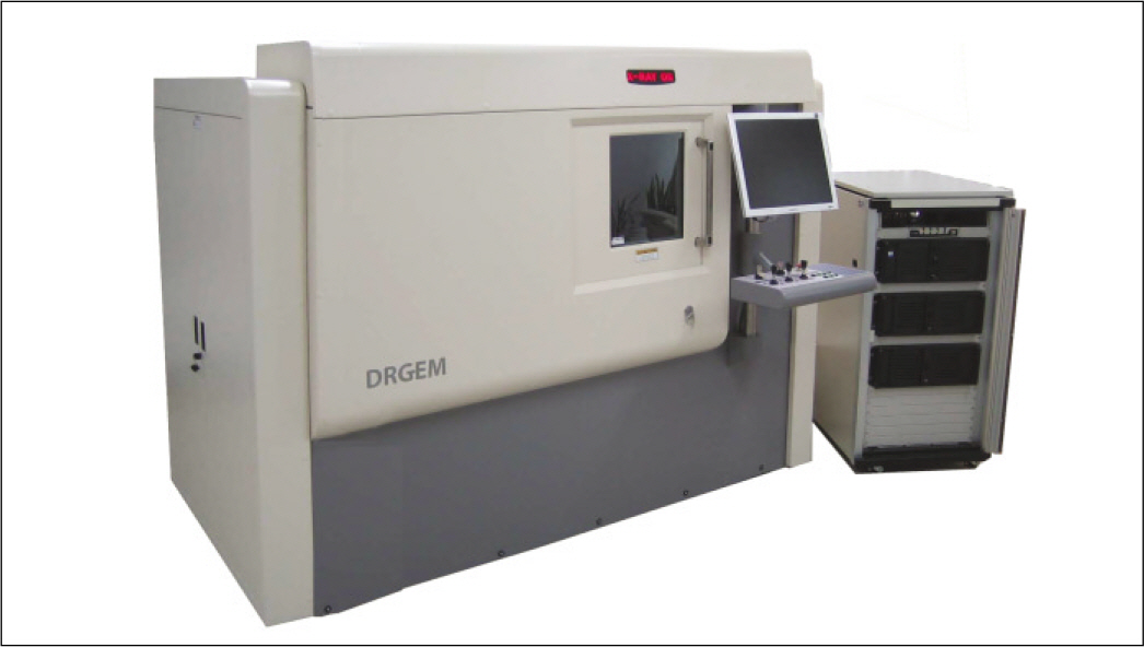
Fig. 1. The microCT used in this study.
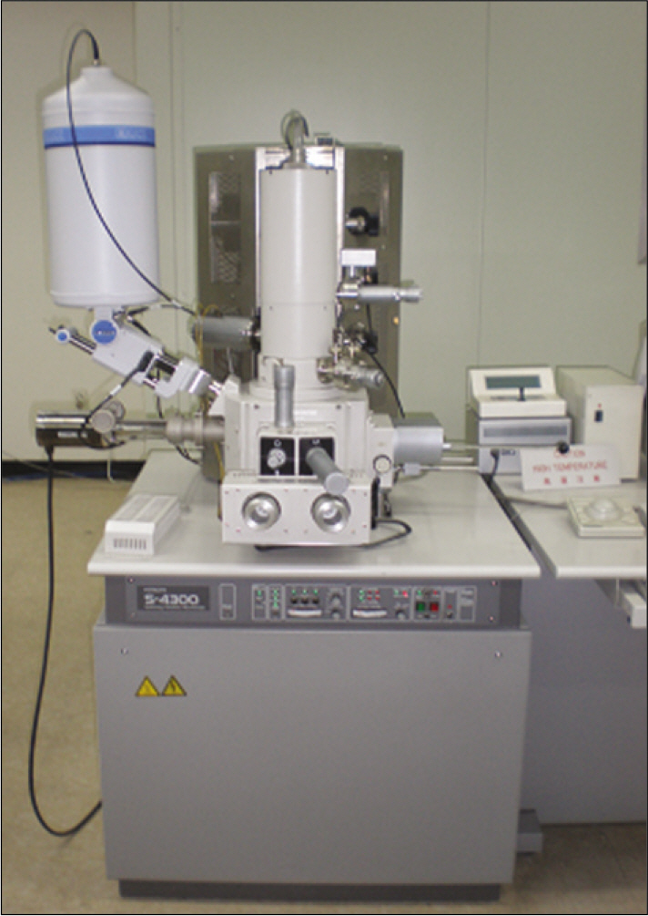
Fig. 2. The FE-SEM used in this study.
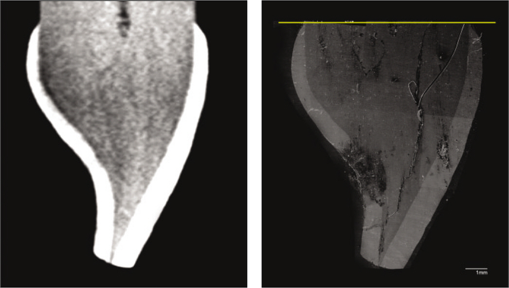
Fig. 3. MicroCT image (left) and scanning electron micrograph (right) of the same cross-section Scale bar = 1 mm.
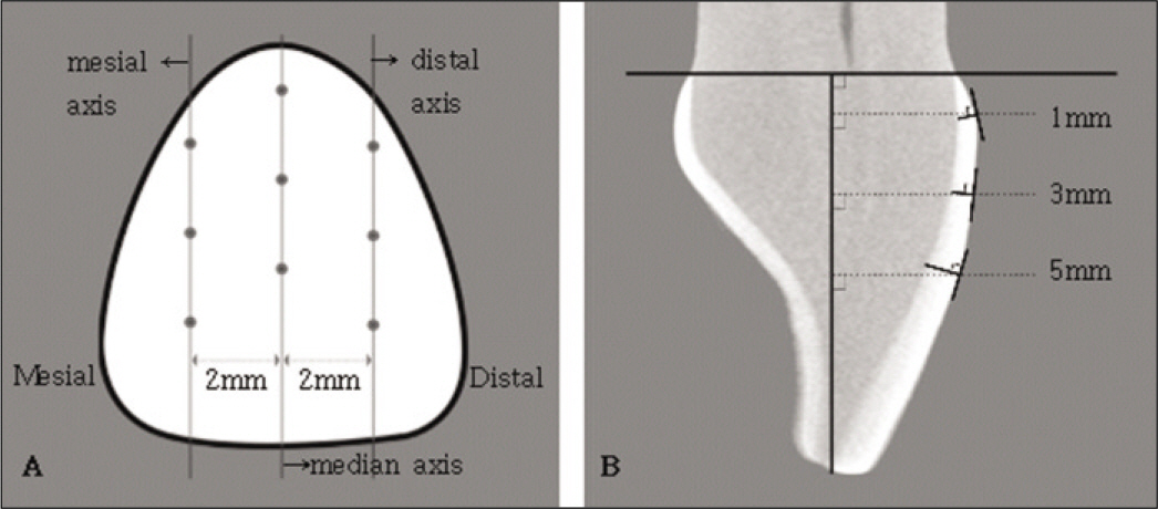
Fig. 4. Tooth landmarks used for measurements A, Labial surface of maxillary incisor. Points are 1, 3 and 5 mm above the CEJ. B, Sagittal plane in the median axis.
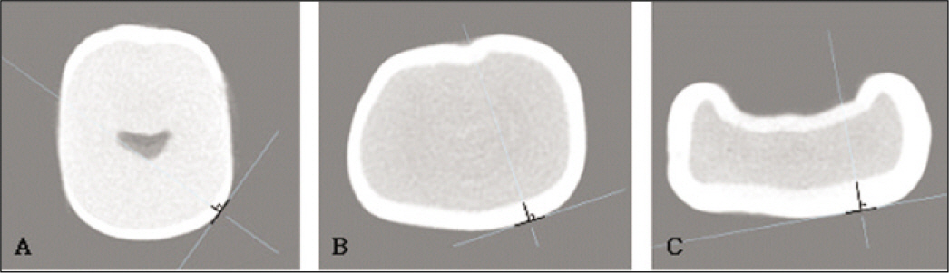
Fig. 5. Method of measurements in 2 mm remote axis mesially and distally from the medial axis A, Horizontal plane 1 mm above the CEJ. B, Horizontal plane 3 mm above the CEJ. C, Horizontal plane 5 mm above the CEJ.
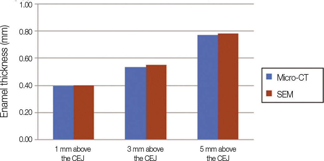
Fig. 6. Comparison of enamel thickness measured with microCT and SEM.
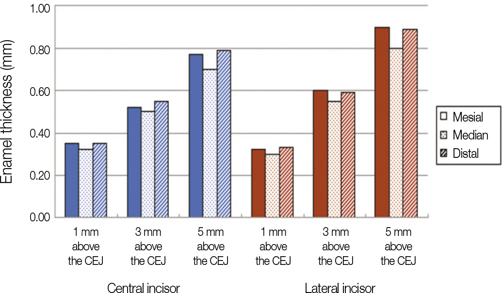
Fig. 7. Comparison of enamel thickness on the each measurement site.
Reference
-
1.Freidman MJ. Augmenting restorative dentistry with porcelain veneers. J Am Dent Assoc. 1991. 122:29–34.2.Shillingburg HT., Hobo S., Whitsett LD., Jacobi R., Brackett SE. Fundamentals of Fixed Prosthodontics. 3rd ed.Chicago: Quintessence;1997. p. 441.3.Christensen GJ. Veneering of teeth. State of the art. Dent Clin North Am. 1985. 29:373–91.4.Clyde JS., Gilmour A. Porcelain veneers: a preliminary review. Br Dent J. 1988. 164:9–14.
Article5.Tjan AH., Dunn JR., Sanderson IR. Microleakage patterns of porcelain and castable ceramic laminate veneers. J Prosthet Dent. 1989. 61:276–82.
Article6.Ferrari M., Patroni S., Balleri P. Measurement of enamel thickness in relation to reduction for etched laminate veneers. Int J Periodontics Restorative Dent. 1992. 12:407–13.7.Edelhoff D., Sorensen JA. Tooth structure removal associated with various preparation designs for anterior teeth. J Prosthet Dent. 2002. 87:503–9.
Article8.Christensen GJ. Has tooth structure been replaced? J Am Dent Assoc. 2002. 133:103–5.
Article9.Walls AW., Steele JG., Wassell RW. Crowns and other extra-coronal restorations: porcelain laminate veneers. Br Dent J. 2002. 193:73–6. 79–82.
Article10.Sheets CG., Taniguchi T. Advantages and limitations in the use of porcelain veneer restorations. J Prosthet Dent. 1990. 64:406–11.
Article11.Hui KK., Williams B., Davis EH., Holt RD. A comparative assessment of the strengths of porcelain veneers for incisor teeth dependent on their design characteristics. Br Dent J. 1991. 171:51–5.
Article12.Kedici PS., Kalipcilar B., Bilir OG. Effect of glass ionomer liners on bonding strength of laminate veneers. J Prosthet Dent. 1992. 68:29–32.
Article13.Peumans M., Van Meerbeek B., Lambrechts P., Vanherle G. Porcelain veneers: a review of the literature. J Dent. 2000. 28:163–77.
Article14.Sorensen JA., Munksgaard EC. Relative gap formation of resin-cemented ceramic inlays and dentin bonding agents. J Prosthet Dent. 1996. 76:374–8.
Article15.Schneider PM., Messer LB., Douglas WH. The effect of enamel surface reduction in vitro on the bonding of composite resin to permanent human enamel. J Dent Res. 1981. 60:895–900.
Article16.Lacy AM., Wada C., Du W., Watanabe L. In vitro microleakage at the gingival margin of porcelain and resin veneers. J Prosthet Dent. 1992. 67:7–10.
Article17.Zaimoglu A., Karaagaçlioglu L. Microleakage in porcelain laminate veneers. J Dent. 1991. 19:369–72.18.Zaimog ̆lu A., Karaag ̆açliog ̆lu L. Uçtaçli. Influence of porcelain ma-S terial and composite luting resin on microleakage of porcelain laminate veneers. J Oral Rehabil. 1992. 19:319–27.19.Sim C., Neo J., Chua EK., Tan BY. The effect of dentin bonding agents on the microleakage of porcelain veneers. Dent Mater. 1994. 10:278–81.
Article20.Atsu SS., Aka PS., Kucukesmen HC., Kiliçarslan MA., Atakan C. Age-related changes in tooth enamel as measured by electron microscopy: implications for porcelain laminate veneers. J Prosthet Dent. 2005. 94:336–41.
Article21.Grine FE., Stevens NJ., Jungers WL. An evaluation of dental radiograph accuracy in the measurement of enamel thickness. Arch Oral Biol. 2001. 46:1117–25.
Article22.Harris EF., Hicks JD. A radiographic assessment of enamel thickness in human maxillary incisors. Arch Oral Biol. 1998. 43:825–31.
Article23.Stroud JL., English J., Buschang PH. Enamel thickness of the posterior dentition: its implications for nonextraction treatment. Angle Orthod. 1998. 68:141–6.24.Stroud JL., Buschang PH., Goaz PW. Sexual dimorphism in mesiodistal dentin and enamel thickness. Dentomaxillofac Radiol. 1994. 23:169–71.
Article25.Scotti R., Villa L., Carossa S. Clinical applicability of the radiographic method for determining the thickness of calcified crown tissues. J Prosthet Dent. 1991. 65:65–7.
Article26.Barber FE., Lees S., Lobene RR. Urasonic pulse-echo measurements in teeth. Arch Oral Biol. 1969. 14:745–60.27.Huysmans MC., Thijssen JM. Ultrasonic measurement of enamel thickness: a tool for monitoring dental erosion? J Dent. 2000. 28:187–91.
Article28.Schwartz GT., Thackeray JF., Reid C., van Reenan JF. Enamel thickness and the topography of the enamel-dentine junction in South African Plio-Pleistocene hominids with special reference to the Carabelli trait. J Hum Evol. 1998. 35:523–42.
Article29.Beynon AD., Wood BA. Variations in enamel thickness and structure in East African hominids. Am J Phys Anthropol. 1986. 70:177–93.
Article30.Olejniczak AJ., Grine FE. Assessment of the accuracy of dental enamel thickness measurements using microfocal X-ray computed tomography. Anat Rec A Discov Mol Cell Evol Biol. 2006. 288:263–75.
Article31.Piemjai M., Arksornnukit M. Compressive fracture resistance of porcelain laminates bonded to enamel or dentin with four adhesive systems. J Prosthodont. 2007. 16:457–64.
Article32.Nakamura T., Miyamae M., Koh N., Hino T., Maruyama T. Adhesive strength between teeth and resin cements for porcelain laminate veneer. J Osaka Univ Dent Sch. 1992. 32:21–6.33.Dumfahrt H., Scha ¨ffer H. Porcelain laminate veneers. A retrospective evaluation after 1 to 10 years of service: Part II-Clinical results. Int J Prosthodont. 2000. 13:9–18.34.Horn HR. Porcelain laminate veneers bonded to etched enamel. Dent Clin North Am. 1983. 27:671–84.35.Calamia JR. Etched porcelain veneers: the current state of the art. Quintessence Int. 1985. 16:5–12.36.Weinberg LA. Tooth preparation for porcelain laminates. N Y State Dent J. 1989. 55:25–8.37.Chpindel P., Cristou M. Tooth preparation and fabrication of porcelain veneers using a double-layer technique. Pract Periodontics Aesthet Dent. 1994. 6:19–28.38.Garber D. Porcelain laminate veneers: ten years later. Part I: Tooth preparation. J Esthet Dent. 1993. 5:56–62.
Article
- Full Text Links
-
- Actions
-
Cited
- CITED
-
- Close
- Share
-
- Similar articles
-
- SEM/EDS Analysis of the Enamel in Mesiodens
- Relationship between mesiodistal width and enamel thickness in mandibular incisors
- Alveolar bone thickness around maxillary central incisors of different inclination assessed with cone-beam computed tomography
- Analysis of the root position of the maxillary incisors in the alveolar bone using cone-beam computed tomography
- Generalized Pulp Stones of Primary Dentition in a Patient with Molar-Incisor Malformation : A Case Report
