J Korean Acad Prosthodont.
2012 Apr;50(2):106-111. 10.4047/jkap.2012.50.2.106.
Surface roughness and Candida albicans adhesion to flexible denture base according to various polishing methods
- Affiliations
-
- 1Department of Prosthodontics and Institute of Oral Bio-Science, Dental School, Chonbuk National University, Jeonju, Korea. skydent@jbnu.ac.kr
- KMID: 2195500
- DOI: http://doi.org/10.4047/jkap.2012.50.2.106
Abstract
- PURPOSE
The purpose of this study was to compare the effect of 3 chairside polishing methods and laboratory polishing methods on surface roughness and C. albicans adhesion of polyamide denture base.
MATERIALS AND METHODS
Using contact profilometer, the surface of polyamide specimens (25x15x2 mm) was studied after conventional polishing without finishing and after chiarside polishing with 2 chiarside polishing kits and chairside-pumice polishing following finishing with tungsten carbide bur. To evaluate the adhesion of C. albicans, C. albicans suspension was overlayed on the test specimen. And the specimens were incubated for 2 hours. Imprint culture method was achieved and counted the colony on the agar plate. Polished polyamide were evaluated using a scanning electron microscope. The statistics were conducted using one-way ANOVA and in case of difference, Scheffe test and Tamhane's T2 test were used.
RESULTS
Surface roughness (Ra) of surfaces polished with 2 chairside polishing kits had higher than conventional polishing and pumice polishing. The highest roughness value was 0.32 +/- 0.10 microm, and the lowest was 0.02 +/- 0.00 microm. The adhesion of C. albicans on the specimens polished with chairside polishing group and pumice polishing group were increased than conventional polishing group (P<.01).
CONCLUSION
Conventional laboratory polishing was found to produce the smoothest surface and the lowest adhesion of C. albicans. Two groups polished with Chairside polishing kits were similar with respect to surface roughness. Surface of the specimen polished with pumice is significantly smoother than 2 chairside polishing groups, but the result of C. albicans adhesion is that group polished with pumice was similar with 2 chairside polishing groups (P>.01).
MeSH Terms
Figure
-

Fig. 1 Profilometer.

Fig. 2 Surface roughness test. *means significant difference at P<.01 statistically.

Fig. 3 SEM image of polished polyamide surfaces. Control group had regular surface. Scratch of SP group was tighter than S group.
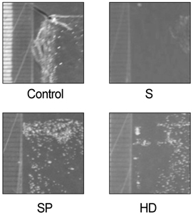
Fig. 4 The colonies of C. albicans on the agar plate.

Fig. 5 The growth of C. albicans on the agar plate. *means significant difference at P<.01 statistically.
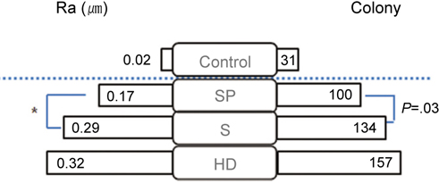
Fig. 6 Comparison of results in this study. *and dotted line mean significantly difference at P<.01 statistically.
Reference
-
1. Renu T, Saurabh G, Samarth KA. Denture base materials: From past to future. Indian J Dent Sci. 2010. 2:33–39.2. Choi B, Kim SH, Lee W. Valplast(R) flexible removable partial denture for a patient with medically compromised conditions : a clinical report. J Korean Acad Prosthodont. 2009. 47:295–299.
Article3. Maurice N. Stern esthetic retention for modern dental prosthesis. NY state Dent J. 1964. 30:53–56.4. Abuzar MA, Bellur S, Duong N, Kim BB, Lu P, Palfreyman N, Surendran D, Tran VT. Evaluating surface roughness of a polyamide denture base material in comparison with poly (methyl methacrylate). J Oral Sci. 2010. 52:577–581.
Article5. Daniluk T, Tokajuk G, Stokowska W, Fiedoruk K, Sciepuk M, Zaremba ML, Rozkiewicz D, Cylwik-Rokicka D, Kedra BA, Anielska I, Górska M, Kedra BR. Occurrence rate of oral Candida albicans in denture wearer patients. Adv Med Sci. 2006. 51:77–80.6. Radford DR, Challacombe SJ, Walter JD. Denture plaque and adherence of Candida albicans to denture-base materials in vivo and in vitro. Crit Rev Oral Biol Med. 1999. 10:99–116.
Article7. Nikawa H, Hamada T, Yamamoto T. Denture plaque-past and recent concerns. J Dent. 1998. 26:299–304.8. Hoepelman IM, Dupont B. Oral candidiasis: the clinical challenge of resistance and management. Int J Antimicrob Agents. 1996. 6:155–159.
Article9. Webb BC, Thomas CJ, Willcox MD, Harty DW, Knox KW. Candida-associated denture stomatitis. Aetiology and management: a review. Part 1. Factors influencing distribution of Candida species in the oral cavity. Aust Dent J. 1998. 43:45–50.
Article10. Takabayashi Y. Characteristics of denture thermoplastic resins for non-metal clasp dentures. Dent Mater J. 2010. 29:353–361.
Article11. Rotrosen D, Calderone RA, Edwards JE Jr. Adherence of Candida species to host tissues and plastic surfaces. Rev Infect Dis. 1986. 8:73–85.
Article12. Bollen CM, Lambrechts P, Quirynen M. Comparison of surface roughness of oral hard materials to the threshold surface roughness for bacterial plaque retention: a review of the literature. Dent Mater. 1997. 13:258–269.
Article13. Quirynen M, Bollen CM. The influence of surface roughness and surface-free energy on supra- and subgingival plaque formation in man. A review of the literature. J Clin Periodontol. 1995. 22:1–14.
Article14. Yamauchi M, Yamamoto K, Wakabayashi M, Kawano J. In vitro adherence of microorganisms to denture base resin with different surface texture. Dent Mater J. 1990. 9:19–24.
Article15. Kuhar M, Funduk N. Effects of polishing techniques on the surface roughness of acrylic denture base resins. J Prosthet Dent. 2005. 93:76–85.
Article16. Radford DR, Challacombe SJ, Walter JD. Denture plaque and adherence of Candida albicans to denture-base materials in vivo and in vitro. Crit Rev Oral Biol Med. 1999. 10:99–116.
Article17. Quirynen M, Bollen CM, Papaioannou W, Van Eldere J, van Steenberghe D. The influence of titanium abutment surface roughness on plaque accumulation and gingivitis: short-term observations. Int J Oral Maxillofac Implants. 1996. 11:169–178.
- Full Text Links
-
- Actions
-
Cited
- CITED
-
- Close
- Share
-
- Similar articles
-
- Prevention of Candida albicans infection in dental polishing lathe by chlorhexidine
- Adherence of Candida to complete denture surfaces in vitro: A comparison of conventional and CAD/CAM complete dentures
- Polishing characteristics of polyetherketoneketone on Candida albicans adhesion
- Biofilm formation on denture base resin including ZnO, CaO, and TiOâ‚‚ nanoparticles
- Evaluation of C. Albicans and S. Mutans adherence on different provisional crown materials
