Screening ultrasonography in pregnancy
- Affiliations
-
- 1Department of Obstetrics and Gynecology, University of Ulsan College of Medicine, Asan Medical Center, Seoul, Korea. hswon@amc.seoul.kr
- KMID: 2195129
- DOI: http://doi.org/10.5124/jkma.2015.58.11.1003
Abstract
- Ultrasonography in obstetrics is increasingly used for the screening of chromosomal abnormalities as well as for prenatal diagnosis of congenital abnormalities with safety and technological advancements. In the first trimester, it is important to confirm normal intrauterine pregnancy with viability, detect the abnormalities of uterus and adnexa, determine the number of fetuses and assess chorionicity and amnionicity in case of multiple pregnancy. After establishment of gestational age accurately by crown-rump length, thickened fetal nuchal translucency, absence of nasal bone, tricuspid regurgitation, reverse a wave of ductus venosus and cystic hygroma can be markers for screening of chromosomal abnormalities. In addition, the scan also offers an opportunity to detect gross structural abnormalities, which could help improve the prognosis by early prenatal intervention. In the second trimester, aneuploidy (trisomy 21, 18, 13, Turner syndrome) and genetic syndromes could be detected by major structural defects and soft markers. It is important to consider that many malformations may not be detected prenatally even by qualified practitioners and appropriate equipment, and to counsel patients about the potential for false-positive or false-negative results.
MeSH Terms
-
Amnion
Aneuploidy
Chorion
Chromosome Aberrations
Congenital Abnormalities
Crown-Rump Length
Female
Fetus
Gestational Age
Humans
Lymphangioma, Cystic
Mass Screening*
Nasal Bone
Nuchal Translucency Measurement
Obstetrics
Pregnancy Trimester, First
Pregnancy Trimester, Second
Pregnancy*
Pregnancy, Multiple
Prenatal Diagnosis
Prognosis
Tricuspid Valve Insufficiency
Trisomy
Ultrasonography*
Ultrasonography, Prenatal
Uterus
Figure
-

Figure 1 Measurement of crown-rump length (arrow) in the mid-sagittal plane of the fetus.
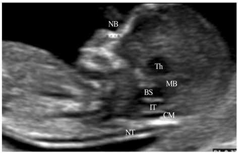
Figure 2 Ultrasound image in the mid-sagittal plane of the fetus showing the nuchal translucency (NT), nasal bone (NB) and intracranial translucency (IT). Th, thalamus; MB, midbrain; BS, brain stem; CM, cisterna magna.
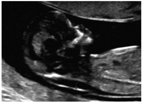
Figure 3 Ultrasound image of the fetus with increased nuchal translucency thickness (3.4 mm) at 12.3 weeks of gestation.
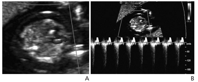
Figure 4 (A) Color and (B) pulsed wave Doppler of tricuspid regurgitation at 12.4 weeks of gestation.

Figure 5 Abnormal Doppler waveforms across the ductus venosus in the parasagittal plane of the fetus at 11.6 weeks of gestation.

Figure 6 Cystic hygroma with generalized skin edema (A) in the sagittal plane and (B) the transverse plane of the fetal head at 12.1 weeks of gestation. CRL, crown-rump length.
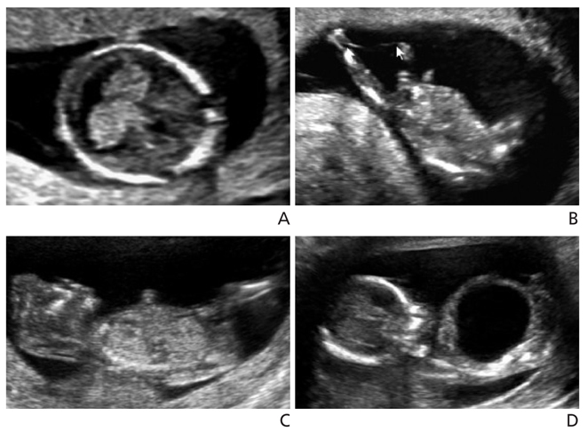
Figure 7 First trimester structural abnormalities. (A) Holoprosencephaly; monoventricle with absent butterfly sign in the transverse plane of the fetal head at 12.5 weeks of gestation. (B) Amniotic band syndrome; thin membranes (arrow) attached to the lower extremities resulted in limb defects at 11.5 weeks of gestation. (C) Acrania; calvarium was absent and remained cranial contents protruded above orbits in the sagittal plane of the fetus at 12.5 weeks of gestation. (D) Bladder outlet obstruction; an enlarged bladder was observed in the male fetus at 13.1 weeks of gestation. Posterior urethral valves was confirmed postnatally and renal function was normal with prenatal vesico-amniotic shunting.

Figure 8 Second trimester sonographic findings in trisomy 21. (A) Absent nasal bone in the mid-sagittal plane of the fetal profile. (B) Cystic hygroma in the transverse plane of the fetal head. (C) Atrioventricular septal defect in the four-chamber view of the fetal heart. (D) Duodenal atresia; the double-bubble sign in the transverse plane of the fetal abdomen.
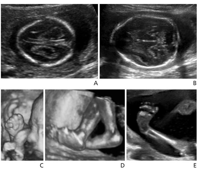
Figure 9 Typical second trimester ultrasonographic findings in trisomy 18. (A) Bilateral choroid plexus cysts and (B) strawberry-shaped skull in the transverse plane of the fetal head. (C) Clenched hands with overlapping fingers and (D) rocker bottom foot in 3D image. (E) Club foot; the plantar surface of the foot was seen in the same plane as the tibia.
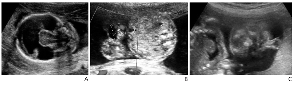
Figure 10 Second trimester structural anomalies in trisomy 13. (A) Holoprosencephaly; monoventricle and fused thalamus in the transverse plane of the fetal head. (B) Omphalocele; the transverse plane of the fetal abdomen showed 11×12mm sized herniated mass into the umbilical cord. (C) Proboscis; coronal image of the face showed protruding tissue above the orbit.
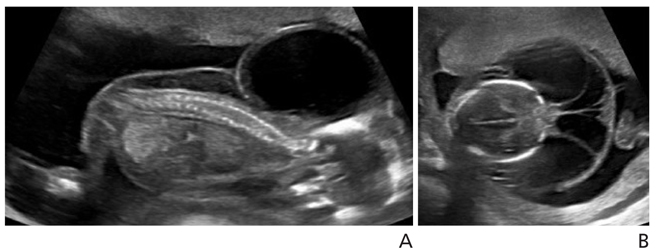
Figure 11 Cystic hygroma with generalized edema in the sagittal plane (A) and the transverse plane (B) of the fetus with Turner syndrome at 16.5 weeks of gestation.
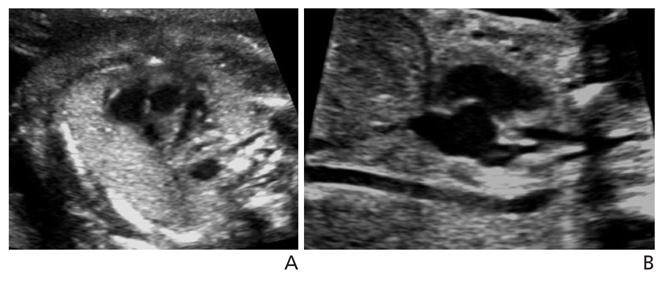
Figure 12 Second trimester ultrasonographic findings in CATCH 22 syndrome. (A) Absent thymus in the 3-vessel view of the fetal heart. (B) Interrupted aortic arch; the ascending aorta was straight and a transverse aortic arch was not demonstrated.
Cited by 1 articles
-
Shift of paradigm in prenatal diagnosis
Do Yeong Hwang
J Korean Med Assoc. 2015;58(11):976-978. doi: 10.5124/jkma.2015.58.11.976.
Reference
-
1. Souka AP, Snijders RJ, Novakov A, Soares W, Nicolaides KH. Defects and syndromes in chromosomally normal fetuses with increased nuchal translucency thickness at 10-14 weeks of ges-tation. Ultrasound Obstet Gynecol. 1998; 11:391–400.
Article2. Snijders RJ, Noble P, Sebire N, Souka A, Nicolaides KH. UK multicentre project on assessment of risk of trisomy 21 by ma-ternal age and fetal nuchal-translucency thickness at 10-14 weeks of gestation. Fetal Medicine Foundation First Trimester Screening Group. Lancet. 1998; 352:343–346.
Article3. Oh SY, Hong JS, Seol HJ, Hwang HS, Park HS, Kim K, Ko HS, Kwak DW, Kim MY, Park MH, Oh MJ, Park JS, Kim SJ. Korean Society of Ultrasound in Obstetrics and Gynecology Research Group. 2014 First-trimester ultrasound forum from the Korean Society of Ultrasound in Obstetrics and Gynecology. Obstet Gynecol Sci. 2015; 58:1–9.
Article4. Sonek J, Nicolaides K. Additional first-trimester ultrasound markers. Clin Lab Med. 2010; 30:573–592.
Article5. Kurjak A, Kupesic S, Matijevic R, Kos M, Marton U. First tri-mester malformation screening. Eur J Obstet Gynecol Reprod Biol. 1999; 85:93–96.
Article6. Cicero S, Curcio P, Papageorghiou A, Sonek J, Nicolaides K. Absence of nasal bone in fetuses with trisomy 21 at 11-14 weeks of gestation: an observational study. Lancet. 2001; 358:1665–1667.
Article7. Kagan KO, Cicero S, Staboulidou I, Wright D, Nicolaides KH. Fetal nasal bone in screening for trisomies 21, 18 and 13 and Turner syndrome at 11-13 weeks of gestation. Ultrasound Obstet Gynecol. 2009; 33:259–264.
Article8. Jung E, Won HS, Lee PR, Kim A. Ultrasonographic measure-ment of fetal nasal bone length in the second trimester in Ko-rean population. Prenat Diagn. 2007; 27:154–157.
Article9. Huggon IC, DeFigueiredo DB, Allan LD. Tricuspid regurgi-tation in the diagnosis of chromosomal anomalies in the fetus at 11-14 weeks of gestation. Heart. 2003; 89:1071–1073.
Article10. Falcon O, Faiola S, Huggon I, Allan L, Nicolaides KH. Fetal tri-cuspid regurgitation at the 11+0 to 13+6-week scan: association with chromosomal defects and reproducibility of the method. Ultrasound Obstet Gynecol. 2006; 27:609–612.
Article11. Faiola S, Tsoi E, Huggon IC, Allan LD, Nicolaides KH. Likeli-hood ratio for trisomy 21 in fetuses with tricuspid regurgitation at the 11 to 13+6-week scan. Ultrasound Obstet Gynecol. 2005; 26:22–27.
Article12. Maiz N, Nicolaides KH. Ductus venosus in the first trimester: contribution to screening of chromosomal, cardiac defects and monochorionic twin complications. Fetal Diagn Ther. 2010; 28:65–71.
Article13. Donnenfeld AE, Lockwood D, Lamb AN. Prenatal diagnosis from cystic hygroma fluid: the value of fluorescence in situ hybridization. Am J Obstet Gynecol. 2001; 185:1004–1008.
Article14. Bornstein E, Goncalves Rodríguez JL, Alvarez Pavon EC, Quiroga H, Or D, Divon MY. First-trimester sonographic find-ings associated with a Dandy-Walker malformation and inferior vermian hypoplasia. J Ultrasound Med. 2013; 32:1863–1868.
Article15. Chaoui R, Benoit B, Mitkowska-Wozniak H, Heling KS, Nicol-aides KH. Assessment of intracranial translucency (IT) in the detection of spina bifida at the 11-13-week scan. Ultrasound Obstet Gynecol. 2009; 34:249–252.
Article16. Lee MY, Won HS, Jeong BD, Hyun MK, Lee HY, Shim JY, Lee PR, Kim A. Measurement of intracranial translucency using three-dimensional ultrasound and Volume IT. Prenat Diagn. 2012; 32:472–475.
Article17. Kim SK, Won HS, Shim JY, Kim KS, Lee PR, Kim A. Successful vesicoamniotic shunting of posterior urethral valves in the first trimester of pregnancy. Ultrasound Obstet Gynecol. 2005; 26:666–668.
Article18. Salomon LJ, Alfirevic Z, Berghella V, Bilardo C, Hernandez-Andrade E, Johnsen SL, Kalache K, Leung KY, Malinger G, Munoz H, Prefumo F, Toi A, Lee W. ISUOG Clinical Standards Committee. Practice guidelines for performance of the routine mid-trimester fetal ultrasound scan. Ultrasound Obstet Gynecol. 2011; 37:116–126.
Article19. Song HK, Ryu HM, Roh SH, Park SH, Kim MY, Kim ES, Han HW, Choi SK, Lee YH, Yoo SJ. Prenatal diagnosis and sono-graphic findings of down syndrome: review of 30 cases. Korean J Obstet Gynecol. 1997; 40:2826–2832.20. Vintzileos AM, Egan JF. Adjusting the risk for trisomy 21 on the basis of second-trimester ultrasonography. Am J Obstet Gynecol. 1995; 172:837–844.
Article21. Yeo L, Vintzileos AM. The second trimester genetic sonogram. In : Callen PW, editor. Ultrasonography in obstetrics and gynecology. 5th ed. London: Elsevier Health Sciences;2011. p. 70–111.22. Nyberg DA, Kramer D, Resta RG, Kapur R, Mahony BS, Luthy DA, Hickok D. Prenatal sonographic findings of trisomy 18: review of 47 cases. J Ultrasound Med. 1993; 12:103–113.
Article23. Cho RC, Chu P, Smith-Bindman R. Second trimester prenatal ultrasound for the detection of pregnancies at increased risk of trisomy 18 based on serum screening. Prenat Diagn. 2009; 29:129–139.
Article24. Song MJ. Fetal sonography for the detection of chromosomal abnormality. J Korean Soc Ultrasound Med. 2011; 30:75–82.25. Papp C, Szigeti Z, Toth-Pal E, Hajdu J, Joo JG, Papp Z. Ultra-sonographic findings of fetal aneuploidies in the second tri-mester: our experiences. Fetal Diagn Ther. 2008; 23:105–113.
Article26. Momma K. Cardiovascular anomalies associated with chromo-some 22q11.2 deletion syndrome. Am J Cardiol. 2010; 105:1617–1624.
Article27. Lee MY, Won HS, Baek JW, Cho JH, Shim JY, Lee PR, Kim A. Variety of prenatally diagnosed congenital heart disease in 22q11.2 deletion syndrome. Obstet Gynecol Sci. 2014; 57:11–16.
Article28. Driscoll DA. Prenatal diagnosis of the 22q11.2 deletion syn-drome. Genet Med. 2001; 3:14–18.
Article
- Full Text Links
-
- Actions
-
Cited
- CITED
-
- Close
- Share
-
- Similar articles
-
- Simple Screening Using Ultrasonography for Prediction of Gestational Diabetes Mellitus
- Medical auditing of whole-breast screening ultrasonography
- Characteristics of breast cancer detected by supplementary screening ultrasonography
- Clinical significance of sonographic soft markers: A review
- Prenatal screening for neural tube defects: from maternal serum alpha-fetoprotein to ultrasonography
