J Korean Neurosurg Soc.
2016 May;59(3):192-196. 10.3340/jkns.2016.59.3.192.
Development and Growth of the Normal Cranial Vault : An Embryologic Review
- Affiliations
-
- 1Department of Neurosurgery, Ansan Hospital, Korea University College of Medicine, Ansan, Korea.
- 2Department of Neurosurgery, Jeju National University Hospital, Jeju, Korea. kibumsim@gmail.com
- KMID: 2192084
- DOI: http://doi.org/10.3340/jkns.2016.59.3.192
Abstract
- Understanding the development of a skull deformity requires an understanding of the normal morphogenesis of the cranium. Craniosynostosis is the premature, pathologic ossification of one or more cranial sutures leading to skull deformities. A review of the English medical literature using textbooks and standard search engines was performed to gather information about the prenatal development and growth of the cranial vault of the neurocranium. A process of morphogenic sequencing begins during prenatal development and growth, continues postnatally, and contributes to the basis for the differential manner of growth of cranial vault bones. This improved knowledge might facilitate comprehension of the pathophysiology of craniosynostosis.
Keyword
MeSH Terms
Figure
-
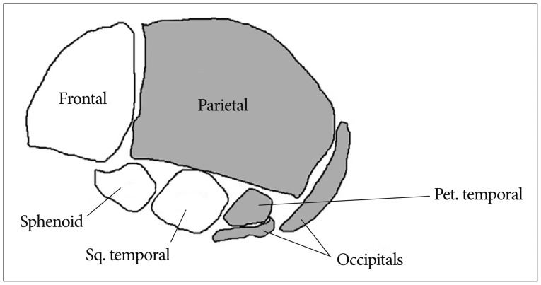
Fig. 1 Skeletal structure of skull : mesenchyme for these structures is derived from neural crest (white) and paraxial mesoderm (gray). Sq : squamous, Pet : petrous.
Cited by 1 articles
-
Preface : Invited Issue Editor Professor Dachling Pang and the Changing Concepts in Spinal Dysraphism during the Last Two Decades
Chae-Yong Kim, Seung-Ki Kim
J Korean Neurosurg Soc. 2020;63(3):269-271. doi: 10.3340/jkns.2020.0064.
Reference
-
1. Adeeb N, Mortazavi MM, Tubbs RS, Cohen-Gadol AA. The cranial dura mater : a review of its history, embryology, and anatomy. Childs Nerv Syst. 2012; 28:827–837. PMID: 22526439.
Article2. Caetano-Lopes J, Canhão H, Fonseca JE. Osteoblasts and bone formation. Acta Reumatol Port. 2007; 32:103–110. PMID: 17572649.3. Cohen MM, MacLean RE. Craniosynostosis : Diagnosis, Evaluation, and Management. ed 2. New York: Oxford University Press;2000. p. 105–107.4. Dixon AD, Hoyte DA, Ronning O. Fundamentals of Craniofacial Growth. New York: CRC Press;1997. p. 101–102.5. Glorieux FH, Pettifor JM, Juppner H. Pediatric bone : Biology & Diseases. San Diego: Academic Press;2003. p. 77–103.6. Hay ED. The mesenchymal cell, its role in the embryo, and the remarkable signaling mechanisms that create it. Dev Dyn. 2005; 233:706–720. PMID: 15937929.
Article7. Liem T. Cranial Osteopathy. Edinburgh, UK: Churchill Livingstone;2005. p. 41–48.8. Moore KL, Persaud TVN, Torchia MG. The Developing Human : Clinically Oriented Embryology. ed 10. Philadelphia: Elsevier Health Sciences;2015. p. 389–390.9. O'Rahilly R, Müller F. Developmental Stages in Human Embryos. Washington D.C.: Carnegie Institution of Washington;1987. p. 1–3.10. Opperman LA. Cranial sutures as intramembranous bone growth sites. Dev Dyn. 2000; 219:472–485. PMID: 11084647.
Article11. Rodeck CH, Whittle MJ. Fetal medicine : Basic Science and Clinical Practice. ed 2. London: Churchill Livingstone;2009. p. 39–41.12. Sadler TW. Langman's Medical Embryology. ed 10. Baltimore, MD: Lippincott Williams & Wilkins;2011. p. 125–127.13. Singh G. Textbook of Orthodontics. ed 3. New Delhi: JP Medical Ltd;2015. p. 44–45.14. Sperber GH, Sperber SM, Guttmann GD. Craniofacial Embryogenetics and Development. ed 2. Shelton, CT: PMPH-USA;2010. p. 95–118.
- Full Text Links
-
- Actions
-
Cited
- CITED
-
- Close
- Share
-
- Similar articles
-
- Sphenoid Bone Determines the Curvature of the Cranial Vault in Postnatal Skull Development in C57BL/6 Mice
- Primary Cranial Vault Lymphoma: A Case Report
- Bull's Osteotomy for Reshaping the Forehead in Simple Symmetric Craniosynostosis
- Primary Mlignant non-Hodgkin's Lymphoma of Cranial Vault
- The Clinical Analysis of 22 Cases of Encephalocele
