A Primary Ossifying Intracranial Myxoma Arising from the Ethmoid Sinus
- Affiliations
-
- 1Department of Neurosurgery, Hanyang University Guri Hospital, Guri, Korea. cjh2324@hanyang.ac.kr
- KMID: 2191384
- DOI: http://doi.org/10.3340/jkns.2015.58.3.281
Abstract
- Myxomas are rare benign tumors that originate from mesenchymal tissue. They usually develop in the atrium of the heart, the skin, subcutaneous tissue, or bone. Involvement of the skull base with an intracranial extension is very rare and not well-described in the literature. We report a rare case of primary intracranial ossifying myxoma arising from the anterior skull base and mimicking a huge chondrosarcoma, and we review the relevant literature.
Keyword
Figure
-
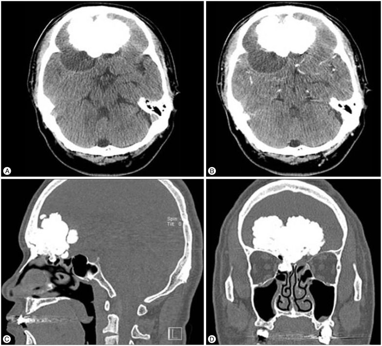
Fig. 1 Preoperative brain (A and B) facial CT (C and D) scans showing giant bony lesions arising from the right ethmoid sinus (C and D), and a low density, multi-lobulated mass with slight peripheral enhancement (A and B).
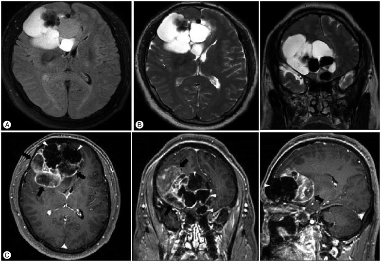
Fig. 2 Preoperative brain magnetic resonance (MR) images. A : Axial Flare MR image showing peripheral high signal lesion and multiple mixed iso- and low signal mass. B : Axial and coronal T2 weighted MR images demonstrating a high signal lesion and central low signal mass. C : Illustrating the giant low-signal lesion (white arrow-heads) and multi-lobulated hypo- or iso-signal mass (black arrows) with mild uneven peripheral gadolinium enhancement.
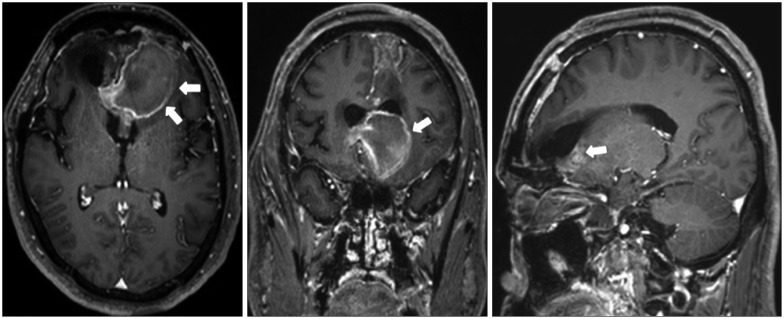
Fig. 3 Postoperative T1-weighted MR images with gadolinium enhancement in the axial, coronal, and sagittal plans show subacute hematoma at left frontal area (white arrows), total resection of the cystic lesion.

Fig. 4 Gross specimens showing the bony structure (left) and jelly-like glistening nature (right) of the resected myxoma.
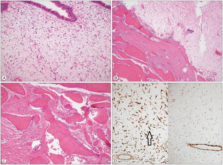
Fig. 5 Light microscopic and immunohistochemical findings. A : Tumor tissue is largely composed of haphazardly scattered stellate cells embedded in the loose myxoid stroma and is continuously lined by pseudostratified respiratory epithelium (H&E stain, ×100). B : Tumor base shows prominent ossification with adjacent fibrous stromal reaction, which is abutting on myxoma area (H&E stain, ×40). C : High power view of ossified fibrotic area (H&E stain, ×100). D : Myxoma cells are positive for Vimentin (Left, Vimentin, ×100, black arrow) but negative for SMA (Right, SMA, ×100). SMA : smooth muscle actin.
Reference
-
1. Andrews T, Kountakis SE, Maillard AA. Myxomas of the head and neck. Am J Otolaryngol. 2000; 21:184–189. PMID: 10834553.
Article2. Branch CL Jr, Laster DW, Kelly DL Jr. Left atrial myxoma with cerebral emboli. Neurosurgery. 1985; 16:675–680. PMID: 4000442.
Article3. DeFatta RJ, Verret DJ, Ducic Y, Carrick K. Giant myxomas of the maxillofacial skeleton and skull base. Otolaryngol Head Neck Surg. 2006; 134:931–935. PMID: 16730532.
Article4. Hsieh DL, Tseng HM, Young YH. Audiovestibular evolution in a patient undergoing surgical resection of a temporal bone myxoma. Eur Arch Otorhinolaryngol. 2006; 263:614–617. PMID: 16612611.
Article5. Lo Muzio L, Nocini P, Favia G, Procaccini M, Mignogna MD. Odontogenic myxoma of the jaws : a clinical, radiologic, immunohistochemical, and ultrastructural study. Oral Surg Oral Med Oral Pathol Oral Radiol Endod. 1996; 82:426–433. PMID: 8899782.6. Nagatani M, Mori S, Takimoto N, Arita N, Ushio Y, Hayakawa T, et al. Primary myxoma in the pituitary fossa : case report. Neurosurgery. 1987; 20:329–331. PMID: 3561744.7. Oruckaptan HH, Sarac S, Gedikoglu G. Primary intracranial myxoma of the lateral skull base : a rare entity in clinical practice. Turk Neurosurg. 2010; 20:86–89. PMID: 20066630.8. Osterdock RJ, Greene S, Mascott CR, Amedee R, Crawford BE. Primary myxoma of the temporal bone in a 17-year-old boy : case report. Neurosurgery. 2001; 48:945–947. discussion 947-948PMID: 11322458.
Article9. Sato H, Gyo K, Tomidokoro Y, Honda N. Myxoma of the sphenoidal sinus. Otolaryngol Head Neck Surg. 2004; 130:378–380. PMID: 15054386.
Article10. Windfuhr JP, Schwerdtfeger FP. Myxoma of the lateral skull base : clinical features and management. Laryngoscope. 2004; 114:249–254. PMID: 14755199.
Article11. Yin H, Cai BW, An HM, You C. Huge primary myxoma of skull base : a report of an uncommon case. Acta Neurochir (Wien). 2007; 149:713–717. PMID: 17558455.
Article12. Zhang L, Zhang M, Zhang J, Luo L, Xu Z, Li G, et al. Myxoma of the cranial base. Surg Neurol. 2007; 68(Suppl 2):S22–S28. PMID: 18037039.
Article13. Zhang LW, Zhang MS, Zhang JT, Luo L, Xu ZL, Li GL, et al. [Myxoma of cranial base : study of 23 cases]. Zhonghua Yi Xue Za Zhi. 2006; 86:1592–1596. PMID: 16854295.
