J Korean Neurosurg Soc.
2014 Jan;55(1):61-63. 10.3340/jkns.2014.55.1.61.
Solitary Cavernous Sinus Neurosarcoidosis Mimicking Neurosyphilis
- Affiliations
-
- 1Department of Neurosurgery, Pusan National University Hospital, Pusan National University School of Medicine, Busan, Korea. mdcwh@naver.com
- 2Department of Otolaryngology, Pusan National University Hospital, Pusan National University School of Medicine, Busan, Korea.
- KMID: 2191045
- DOI: http://doi.org/10.3340/jkns.2014.55.1.61
Abstract
- A differential diagnosis between neurosarcoidosis and neurosyphilis is particularly problematic in patients with a positive serologic result for syphilis. We report here a patient with a solitary cavernous sinus sarcoidosis who had a history of syphilis and showed rapidly progressing cavernous sinus syndrome. A transsphenoidal biopsy was performed and a histopathologic examination revealed a non-caseating granuloma with an asteroid body. His facial pain disappeared after steroid therapy. He received oral prednisolone for one year. A follow-up magnetic resonance imaging of the brain revealed resolution of the mass over the cavernous sinus. Particularly in patients with a history of syphilis, neurosyphilis should be included in a differential diagnosis of neurosarcoidosis.
Keyword
MeSH Terms
Figure
-
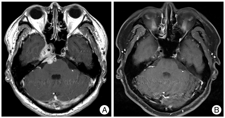
Fig. 1 Gadolinium-enhanced magnetic resonance (MR) images. A : Preoperative MR image revealing the mass extended toward the middle fossa laterally and to the sphenoid sinus anteriorly. This lesion also extended to the cerebellopontine angle posteriorly with a compression of the brain stem. B : Postoperative MR image revealing resolution of the mass over the cavernous sinus region sinus.
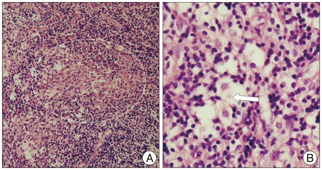
Fig. 2 Photomicrographs of the lesion showing a non-caseating granuloma (circle, A) with an asteroid body (arrow, B). H&E, original magnification, ×100 (A), ×400 (B).
Reference
-
1. Cain H, Kraus B. Asteroid bodies : derivatives of the cytosphere. An electron microscopic contribution to the pathology of the cytocentre. Virchows Arch B Cell Pathol. 1977; 26:119–132. PMID: 204105.2. Carlson JA, Dabiri G, Cribier B, Sell S. The immunopathobiology of syphilis : the manifestations and course of syphilis are determined by the level of delayed-type hypersensitivity. Am J Dermatopathol. 2011; 33:433–460. PMID: 21694502.
Article3. Edmonds LC, Stubbs SE, Ryu JH. Syphilis : a disease to exclude in diagnosing sarcoidosis. Mayo Clin Proc. 1992; 67:37–41. PMID: 1732690.4. Elias WJ, Lanzino G, Reitmeyer M, Jane JA. Solitary sarcoid granuloma of the cerebellopontine angle : a case report. Surg Neurol. 1999; 51:185–190. PMID: 10029426.5. Gascón-Bayarri J, Mañá J, Martínez-Yélamos S, Murillo O, Reñé R, Rubio F. Neurosarcoidosis : report of 30 cases and a literature survey. Eur J Intern Med. 2011; 22:e125–e132. PMID: 22075297.6. Harding AS, Ghanem KG. The performance of cerebrospinal fluid treponemal-specific antibody tests in neurosyphilis : a systematic review. Sex Transm Dis. 2012; 39:291–297. PMID: 22421696.
Article7. Hervier B, Wastiaux H, Freour T, Masseau A, Corvec S, Armingeat T, et al. [Sarcoidosis-like granulomatosis revealing a tertiary syphilis]. Rev Med Interne. 2009; 30:806–808. PMID: 19249139.8. Hoitsma E, Faber CG, Drent M, Sharma OP. Neurosarcoidosis : a clinical dilemma. Lancet Neurol. 2004; 3:397–407. PMID: 15207796.9. Hoitsma E, Sharma OP. Neurosarcoidosis. Eur Respir Mon. 2005; 32:164–187.
Article10. Khoury J, Wellik KE, Demaerschalk BM, Wingerchuk DM. Cerebrospinal fluid angiotensin-converting enzyme for diagnosis of central nervous system sarcoidosis. Neurologist. 2009; 15:108–111. PMID: 19276791.
Article11. Kumar G, Kang CA, Giannini C. Neurosarcoidosis presenting as a cerebellar mass. J Gen Intern Med. 2007; 22:1373–1376. PMID: 17619108.
Article12. Marra CM, Tantalo LC, Maxwell CL, Ho EL, Sahi SK, Jones T. The rapid plasma reagin test cannot replace the venereal disease research laboratory test for neurosyphilis diagnosis. Sex Transm Dis. 2012; 39:453–457. PMID: 22592831.
Article13. Perry HO, Lofgren RK. Secondary and tertiary syphilis presenting as sarcoidal reactions of the skin. Cutis. 1984; 34:253–255. 257–258. PMID: 6488885.14. Rolfes DB, Weiss MA, Sanders MA. Necrotizing sarcoid granulomatosis with suppurative features. Am J Clin Pathol. 1984; 82:602–607. PMID: 6093497.
Article15. Singh R, Kaur D, Parameswaran M. Sarcoidal reaction of the skin in syphilis. Br J Vener Dis. 1971; 47:209–211. PMID: 5090748.
Article16. Spector WG, Heesom N. The production of granulomata by antigen-antibody complexes. J Pathol. 1969; 98:31–39. PMID: 4900707.
Article17. Spencer TS, Campellone JV, Maldonado I, Huang N, Usmani Q, Reginato AJ. Clinical and magnetic resonance imaging manifestations of neurosarcoidosis. Semin Arthritis Rheum. 2005; 34:649–661. PMID: 15692958.
Article18. Stübgen JP. Neurosarcoidosis presenting as a retroclival mass. Surg Neurol. 1995; 43:85–87. discussion 87-88. PMID: 7701433.
Article19. Tanabe JL, Huntley AC. Granulomatous tertiary syphilis. J Am Acad Dermatol. 1986; (2 Pt 2):15:341–344. PMID: 3734178.
Article20. Terushkin V, Stern BJ, Judson MA, Hagiwara M, Pramanik B, Sanchez M, et al. Neurosarcoidosis: presentations and management. Neurologist. 2010; 16:2–15. PMID: 20065791.
