Pure Intramuscular Osteolipoma
- Affiliations
-
- 1Department of Neurosurgery, Spine Center, Chuncheon Sacred Heart Hospital, College of Medicine, Hallym University, Chuncheon, Korea. nscharisma@hanmail.net
- KMID: 2190950
- DOI: http://doi.org/10.3340/jkns.2013.54.6.518
Abstract
- Ossified lipoma or osteolipoma are rarely reported. It is defined as a histologic variant of lipoma that has undergone osseous metaplasia. Osteolipoma presents with a dominant osseous component within a lipoma. We report a case of a histologically confirmed osteolipoma on the nuchal ligament independent of bone. The patient was a 51-year-old female who presented with a 5-year history of a painless, progressively enlarging mass on the posterior neck. Computed tomography and magnetic resonance imaging showed a circumscribed mass compatible with fat between the C2 and C6 spinous processes with a large calcified irregular component. The mass with dual components was totally removed under general anesthesia and no recurrence was observed after 6 months of follow-up. We also reviewed the clinicopathologic features of previously reported osteolipomas in the literature and suggest that although osteolipoma is a rare variant of lipoma, it should be considered in the differential diagnosis when a lipoma of the posterior neck mixed with a bony component is encountered.
Keyword
MeSH Terms
Figure
-

Fig. 1 Preoperatively CT (A: sagittal) and sagittal MRI (B: T1-weighted, C: T2-weighted) revealing a irregular calcification involving posterior neck within soft tissue mass compatible with fat tissue.
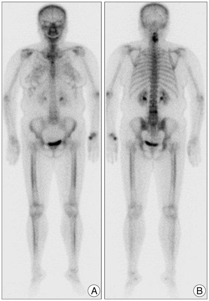
Fig. 2 Whole body bone scan (A: anterior, B: posterior) shows an amorphous calcification mass with increased uptake, and no bone metastases.

Fig. 3 Grossly, the tumor consists largely of fat and calcification presented with the red bone marrow (black arrow).
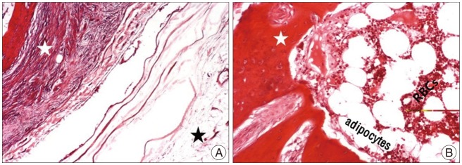
Fig. 4 There is a large foci of osseous metaplasia (white star) and at the periphery of the mass, the bony portion is surrounded by mature adipose tissue (black star) [A: hematoxylin & eosin (H-E), ×40]. Microscopic appearance of red bone marrow shows a meshwork of bone trabeculae (white star) and hematopoietic marrow elements (B: H-E, ×100).
Reference
-
1. Abdalla WM, da Motta AC, Lin SY, McCarthy EF, Zinreich SJ. Intraosseous lipoma of the left frontoethmoidal sinuses and nasal cavity. AJNR Am J Neuroradiol. 2007; 28:615–617. PMID: 17416808.2. Adebiyi KE, Ugboko VI, Maaji SM, Ndubuizu G. Osteolipoma of the palate: report of a case and review of the literature. Niger J Clin Pract. 2011; 14:242–244. PMID: 21860148.
Article3. Bohm KC, Birman MV, Silva SR, Lesperance MM, Marentette LJ, Beyer GR, et al. Ossifying lipoma of c1-c2 in an adolescent. J Pediatr Orthop. 2011; 31:e53–e56. PMID: 21654449.
Article4. Brones A, Mengshol S, Wilkinson CC. Ossifying lipoma of the cervical spine. J Neurosurg Pediatr. 2010; 5:283–284. PMID: 20192646.
Article5. de Castro AL, de Castro EV, Felipini RC, Ribeiro AC, Soubhia AM. Osteolipoma of the buccal mucosa. Med Oral Patol Oral Cir Bucal. 2010; 15:e347–e349. PMID: 19767701.6. Demiralp B, Alderete JF, Kose O, Ozcan A, Cicek I, Basbozkurt M. Osteolipoma independent of bone tissue: a case report. Cases J. 2009; 2:8711. PMID: 19918398.7. Demirkan F, Demirkan N. Ossifying lipoma at the wrist. Aegean Pathol J. 2004; 1:19–21.8. Fritchie KJ, Renner JB, Rao KW, Esther RJ. Osteolipoma: radiological, pathological, and cytogenetic analysis of three cases. Skeletal Radiol. 2012; 41:237–244. PMID: 21822651.
Article9. Gaskin CM, Helms CA. Lipomas, lipoma variants, and well-differentiated liposarcomas (atypical lipomas): results of MRI evaluations of 126 consecutive fatty masses. AJR Am J Roentgenol. 2004; 182:733–739. PMID: 14975977.
Article10. Gokul S, Ranjini KV, Kirankumar K, Hallikeri K. Congenital osteolipoma associated with cleft palate: a case report. Int J Oral Maxillofac Surg. 2009; 38:91–93. PMID: 18952409.
Article11. Heffernan EJ, Lefaivre K, Munk PL, Nielsen TO, Masri BA. Ossifying lipoma of the thigh. Br J Radiol. 2008; 81:e207–e210. PMID: 18628326.
Article12. Jaiswal AK, Garg A, Mahapatra AK. Spinal ossifying lipoma. J Clin Neurosci. 2005; 12:714–717. PMID: 16098751.
Article13. Jamshidi K, Qomashi I, Shirazi MR. An unusual location of ossified intramuscular lipoma: a case report. Acta Med Iran. 2011; 49:630–632. PMID: 22052147.14. Lin YC, Huang CC, Chen HJ. Intraspinal osteolipoma. Case report. J Neurosurg. 2001; 94(1 Suppl):126–128. PMID: 11147847.15. Park YS, Kwon JT, Park US. Interhemispheric osteolipoma with agenesis of the corpus callosum. J Korean Neurosurg Soc. 2010; 47:148–150. PMID: 20224717.
Article16. Plaut GS, Salm R, Truscott DE. Three cases of ossifying lipoma. J Pathol Bacteriol. 1959; 78:292–295. PMID: 14433447.
Article
