Urgent Intracranial Carotid Artery Decompression after Penetrating Head Injury
- Affiliations
-
- 1Department of Neurosurgery, College of Medicine, The Catholic University of Korea, Bucheon St. Mary's Hospital, Bucheon, Korea. ispahk@yahoo.co.kr
- KMID: 2190757
- DOI: http://doi.org/10.3340/jkns.2013.53.3.180
Abstract
- We describe a case of intracranial carotid artery occlusion due to penetrating craniofacial injury by high velocity foreign body that was relieved by decompressive surgery. A 46-year-old man presented with a penetrating wound to his face. A piece of an electric angular grinder disc became lodged in the anterior skull base. Computed tomography revealed that the disc had penetrated the unilateral paraclinoid and suprasellar areas without flow of the intracranial carotid artery on the lesion side. The cavernous sinus was also compromised. Removal of the anterior clinoid process reopened the carotid blood flow, and the injection of glue into the cavernous sinus restored complete hemostasis during extraction of the fragment from the face. Digital subtraction angiography revealed complete recanalization of the carotid artery without any evidence of dissection. Accurate diagnosis regarding the extent of the compromised structures and urgent decompressive surgery with adequate hemostasis minimized the severity of penetrating damage in our patient.
MeSH Terms
Figure
-

Fig. 1 Photograph and CTs obtained at the time of admission. Deep penetrating laceration is visible (A). A piece of grinder blade is visible on facial bone three dimensional CT (B). Preoperative facial bone CTs show the broken blade involving the right cavernous sinus, optic nerve and paraclinoid segment of the internal carotid artery (C and D). Preoperative brain CTs show the involvement of the right temporal lobe, cavernous sinus, paraclinoid segment of the carotid artery (E), and traumatic subarachnoid hemorrhage, pneumocephalus without apparent low attenuation area of the right intracranial carotid artery (F).
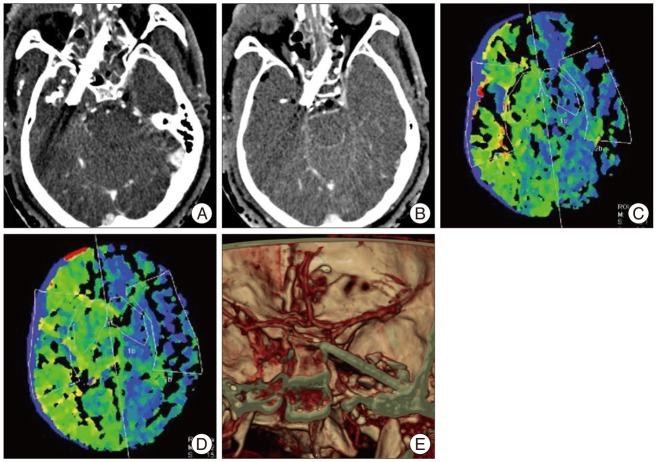
Fig. 2 CT angiography and perfusion study showing the injury of the right internal carotid artery (ICA). Right ICA is not visible while the left ICA density remained visible on the raw data of CT angiography (A and B). Perfusion CTs show the delayed time to peak of the right hemisphere suggesting the right ICA occlusion (C and D). Preoperative CT angiography shows the right ICA impinged between broken blade and anterior clinoid process (E).
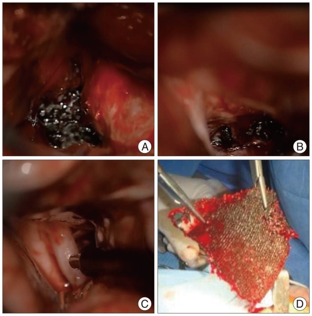
Fig. 3 Operative microscopic view. Extradural view shows the blade fragment penetrating temporal lobe after removal of the anterior clinoid process (A). Intradural views show the displacement of the internal carotid artery (ICA) due to blade fragment (B) and restoration of flow to the ICA after extraction of broken blade (C). The broken blade with the size of 9×10 cm was extracted from the face (D).
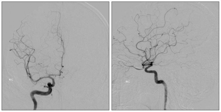
Fig. 4 Postoperative angiography obtained at 2 months after surgery. Complete recanalization of the right internal carotid artery is visible.
Reference
-
1. Abdoli A, Amirjamshidi A. Work-related penetrating head trauma caused by industrial grinder tool. Arch Iran Med. 2009; 12:496–498. PMID: 19722774.2. Back DL, Espag M, Hilton A, Peckham T. Angle grinder injuries. Injury. 2000; 31:475–476. PMID: 10831752.
Article3. Larsen DW. Traumatic vascular injuries and their management. Neuroimaging Clin N Am. 2002; 12:249–269. PMID: 12391635.
Article4. Maier H, Tisch M, Lorenz KJ, Danz B, Schramm A. [Penetrating injuries in the face and neck region. Diagnosis and treatment]. HNO. 2011; 59:765–782. PMID: 21732148.5. Park IH, Kim HS, Park SK, Kim SW. Traumatic pseudoaneurysm of the superficial temporal artery diagnosed by 3-dimensional CT angiography. J Korean Neurosurg Soc. 2008; 43:209–211. PMID: 19096647.
Article6. Sekhar LN, Natarajan SK, Manning T, Bhagawati D. The use of fibrin glue to stop venous bleeding in the epidural space, vertebral venous plexus, and anterior cavernous sinus : technical note. Neurosurgery. 2007; 61(3 Suppl):E51. discussion E51. PMID: 17876220.
- Full Text Links
-
- Actions
-
Cited
- CITED
-
- Close
- Share
-
- Similar articles
-
- A Case of the Common Carotid Artery Penetrating Injury by Grass Cutter Fragment
- Intracranial Occlusion of Internal Carotid Artery in Acute Closed Head Injury: Case Report
- Penetrating Carotid Artery Injuries Treated by an Urgent Endovascular Stent Technique: Report of Two Cases
- A Case of the Zone III Penetrating Neck Injury with Internal Carotid Artery Laceration Treated by an Urgent Endovascular Stent Technique
- A Case of Traumatic Cerebral Infarction due to Injury of the Petrosal Internal Carotid Artery Resulting from Head Trauma
