Combined Open Door Laminoplasty with Unilateral Screw Fixation for Unstable Multi-Level Cervical Stenosis : A Preliminary Report
- Affiliations
-
- 1Department of Neurosurgery, Gachon University, Gil Hospital, Incheon, Korea. samddal@gilhospital.com
- KMID: 2190683
- DOI: http://doi.org/10.3340/jkns.2013.53.2.83
Abstract
OBJECTIVE
The authors reviewed their experiences of combined surgery (open door laminoplasty with unilateral screw fixation) for unstable multi-level cervical stenosis, to clarify the situation regarding the surgical approach most appropriate for the treatment of diffuse unstable multi-level cervical stenosis.
METHODS
From January 2011 to January 2012, combined surgery was performed for unstable multi-level cervical stenosis by one surgeon at our institution. The subjects of this study were 6 men of mean age 53.7 years (range, 48-71) with a mean follow-up of 9.3 (range, 3-14) months. All imaging studies showed severe multi-level cervical stenosis with spinal cord signal change, and instability or kyphotic deformity. A retrospective review of clinical, radiological, and surgical data was conducted.
RESULTS
Average laminoplasty level was 4.8 and the average screw fixation level was 5.0. Japanese Orthopedic Association score improved from an average of 5.2 to 11.2 points. According to Nurick's grades and Odom's criteria, symptom improvement was statistically significant. On the other hand, Cobb's angle changes were not significant. Average operation time was 5.86 hours with an average blood loss of 460 mL. No significant surgical complication was encountered.
CONCLUSION
Despite the small cohort and the short follow-up duration, the present study demonstrates that laminoplasty with unilateral screw fixation is a safe and effective treatment for unstable multi-level cervical stenosis.
MeSH Terms
Figure
-
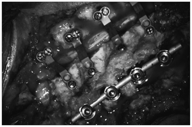
Fig. 1 Intraoperative picture showing combined open door laminoplasty and unilateral screw fixation.
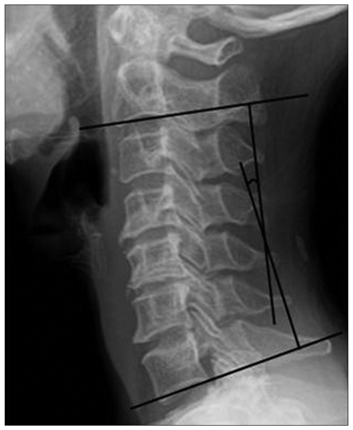
Fig. 2 Cobb's method for measuring cervical lordosis. Cervical lordotic angles are measured by joining perpendiculars to lines drawn parallel to the inferior end plates of C2 and C7. When the C7 vertebra was not well visualized on lateral film, the inferior plate of C6 was used.
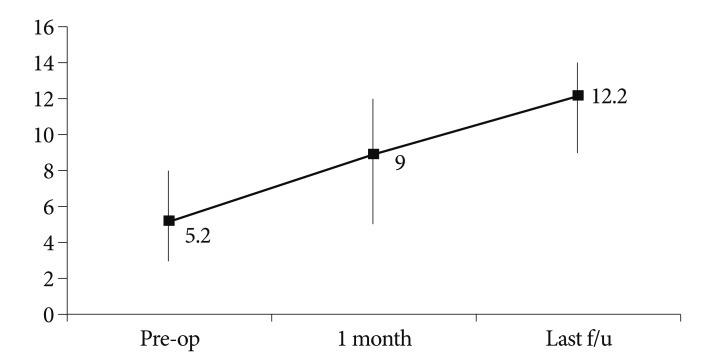
Fig. 3 Graph showing JOA scores before surgery and during follow-up. JOA scores recovered by a mean of 6.0 points (from 5.2 to 11.2; p<0.05) at last follow-up. f/u : follow-up, JOA : Japanese Orthopedic Association, pre-op : pre-operation.

Fig. 4 Graph showing Nurick's grades before surgery and during follow-up. Nurick' s grade recovered by a mean of 2.3 (from 4.5 to 1.8; p<0.05) at last follow-up. f/u : follow-up, pre-op : pre-operation.
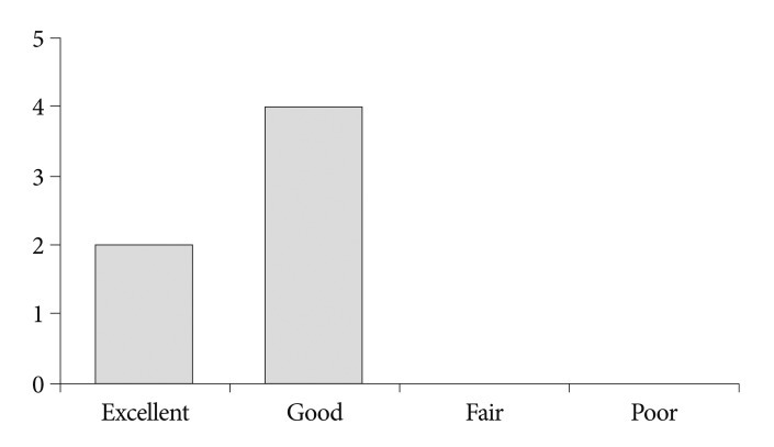
Fig. 5 Graph showing Odom's criteria at last follow-up. The clinical success rate according to Odom's criteria was 100%.

Fig. 6 Graph showing Cobb's angle before surgery and during follow-up. The improvements observed at last follow-up visits were not significant. f/u : follow-up, pre-op : pre-operation.
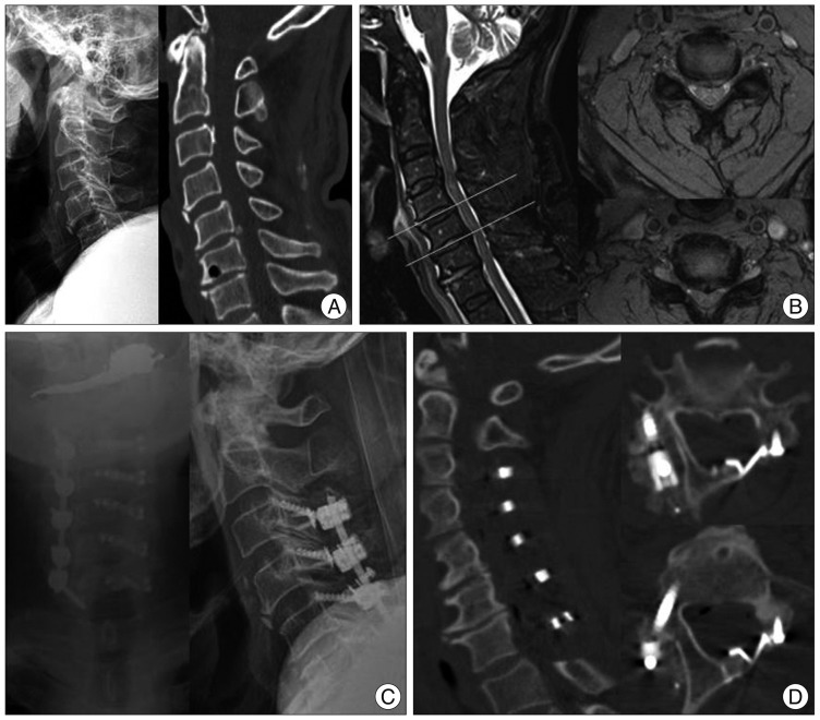
Fig. 7 A 71-year-old man presented with quadriparesis after a slip down. A : Preoperative cervical radiography and CT showed cervical degenerative spondylosis with loss of cervical lordosis and a teardrop fracture of the C4 body with spondylolisthesis at C4-C5 and C5-C6. B : Preoperative cervical MRI demonstrated severe spinal cord compression at C3-C6 with high signal change in T2-weighted images. We suspected an ALL injury based on the signal change observed in pre-vertebral soft tissue. C : Left side open door laminoplasty at C3-C7 with lateral mass screw fixation at C3-C6 and transpedicular screw fixation at C7 were performed. D : Postoperative cervical CT showed an efficiently expanded spinal canal and corrected cervical alignment. ALL : anterior longitudinal ligament, CT : computed tomography, MRI : magnetic resonance image.
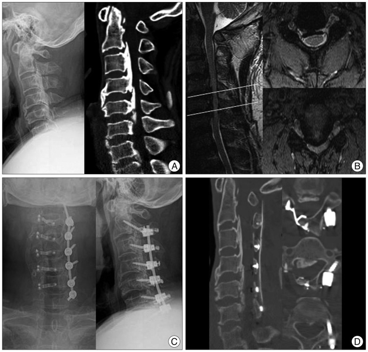
Fig. 8 A 50-year-old man presented with a radiating pain in both arms and progressive gait disturbance. A : Preoperative cervical radiography and CT revealed degenerative spondylosis, loss of lordosis, and diffuse OPLL at C2-C6. B : Preoperative cervical MRI revealed severe spinal cord compression at C2-C6 with high signal change on T2 weighted images. C : Right side open door laminoplasty at C2-C6 with lateral mass screw fixation at C3-C6 and transpedicular screw fixation at C2 and C7 were performed. D : Postoperative cervical CT showed an efficiently expanded spinal canal and appropriately inserted screws. OPLL : ossification of posterior longitudinal ligament, CT : computed tomography, MRI : magnetic resonance image.
Reference
-
1. Albert TJ, Vacarro A. Postlaminectomy kyphosis. Spine (Phila Pa 1976). 1998; 23:2738–2745. PMID: 9879099.
Article2. Chen HC, Chang MC, Yu WK, Wang ST, Liu CL, Chen TH. Lateral mass anchoring screws for cervical laminoplasty : preliminary report of a novel technique. J Spinal Disord Tech. 2008; 21:387–392. PMID: 18679091.3. Chiba K, Ogawa Y, Ishii K, Takaishi H, Nakamura M, Maruiwa H, et al. Long-term results of expansive open-door laminoplasty for cervical myelopathy--average 14-year follow-up study. Spine (Phila Pa 1976). 2006; 31:2998–3005. PMID: 17172996.
Article4. Herkowitz HN. A comparison of anterior cervical fusion, cervical laminectomy, and cervical laminoplasty for the surgical management of multiple level spondylotic radiculopathy. Spine (Phila Pa 1976). 1988; 13:774–780. PMID: 3194786.
Article5. Herkowitz HN. Cervical laminaplasty : its role in the treatment of cervical radiculopathy. J Spinal Disord. 1988; 1:179–188. PMID: 2856537.6. Hirabayashi K, Satomi K. Operative procedure and results of expansive open-door laminoplasty. Spine (Phila Pa 1976). 1988; 13:870–876. PMID: 3143157.
Article7. Hirabayashi K, Toyama Y, Chiba K. Expansive laminoplasty for myelopathy in ossification of the longitudinal ligament. Clin Orthop Relat Res. 1999; 35–48. PMID: 10078127.
Article8. Houten JK, Cooper PR. Laminectomy and posterior cervical plating for multilevel cervical spondylotic myelopathy and ossification of the posterior longitudinal ligament : effects on cervical alignment, spinal cord compression, and neurological outcome. Neurosurgery. 2003; 52:1081–1087. discussion 1087-1088. PMID: 12699550.
Article9. Iwasaki M, Okuda S, Miyauchi A, Sakaura H, Mukai Y, Yonenobu K, et al. Surgical strategy for cervical myelopathy due to ossification of the posterior longitudinal ligament : Part 1 : clinical results and limitations of laminoplasty. Spine (Phila Pa 1976). 2007; 32:647–653. PMID: 17413469.
Article10. Jeanneret B, Magerl F, Ward EH, Ward JC. Posterior stabilization of the cervical spine with hook plates. Spine (Phila Pa 1976). 1991; 16(3 Suppl):S56–S63. PMID: 2028342.
Article11. Kamikozuru M. [Significance of the anterior floating method for cervical myelopathy due to the ossification of the posterior longitudinal ligament]. Nihon Seikeigeka Gakkai Zasshi. 1991; 65:431–440. PMID: 1955790.12. Kawai S, Sunago K, Doi K, Saika M, Taguchi T. Cervical laminoplasty (Hattori's method). Procedure and follow-up results. Spine (Phila Pa 1976). 1988; 13:1245–1250. PMID: 3144758.13. Kim JG, Kim SW, Lee SM, Shin H. Surgical result of the combined anterior and posterior approach in treatment of cervical spondylotic myelopathy. J Korean Neurosurg Soc. 2006; 39:188–191.14. Kim YS, Chin DK, Cho YE, Jin BH, Yoon YS, Park JP, et al. Surgical treatment for ossification of the posterior longitudinal ligament of the cervical spine. J Korean Neurosurg Soc. 1997; 26:1237–1245.15. Kumar VG, Rea GL, Mervis LJ, McGregor JM. Cervical spondylotic myelopathy : functional and radiographic long-term outcome after laminectomy and posterior fusion. Neurosurgery. 1999; 44:771–777. discussion 777-778. PMID: 10201302.
Article16. Lee TT, Green BA, Gromelski EB. Safety and stability of open-door cervical expansive laminoplasty. J Spinal Disord. 1998; 11:12–15. PMID: 9493764.
Article17. Matsumura A, Yanaka K, Akutsu H, Noguchi S, Moritake T, Nose T. Combined laminoplasty with posterior lateral mass plate for unstable spondylotic cervical canal stenosis--technical note. Neurol Med Chir (Tokyo). 2003; 43:514–519. discussion 519. PMID: 14620206.
Article18. Matsunaga S, Sakou T, Nakanisi K. Analysis of the cervical spine alignment following laminoplasty and laminectomy. Spinal Cord. 1999; 37:20–24. PMID: 10025690.
Article19. Mizuno J, Nakagawa H. Ossified posterior longitudinal ligament : management strategies and outcomes. Spine J. 2006; 6(6 Suppl):282S–288S. PMID: 17097548.20. Morimoto T, Yamada T, Okumura Y, Kakizaki T, Kawaguchi S, Hiramatsu K, et al. Expanding laminoplasty for cervical myelopathy-spinous process roofing technique. Acta Neurochir (Wien). 1996; 138:720–725. PMID: 8836288.
Article21. Mummaneni PV, Haid RW, Rodts GE Jr. Combined ventral and dorsal surgery for myelopathy and myeloradiculopathy. Neurosurgery. 2007; 60(1 Suppl 1):S82–S89. PMID: 17204891.
Article22. Nakano K, Harata S, Suetsuna F, Araki T, Itoh J. Spinous process-splitting laminoplasty using hydroxyapatite spinous process spacer. Spine (Phila Pa 1976). 1992; 17(3 Suppl):S41–S43. PMID: 1314432.
Article23. Nakase H, Park YS, Kimura H, Sakaki T, Morimoto T. Complications and long-term follow-up results in titanium mesh cage reconstruction after cervical corpectomy. J Spinal Disord Tech. 2006; 19:353–357. PMID: 16826008.
Article24. Satomi K, Nishu Y, Kohno T, Hirabayashi K. Long-term follow-up studies of open-door expansive laminoplasty for cervical stenotic myelopathy. Spine (Phila Pa 1976). 1994; 19:507–510. PMID: 8184342.
Article25. Satomi K, Ogawa J, Ishii Y, Hirabayashi K. Short-term complications and long-term results of expansive open-door laminoplasty for cervical stenotic myelopathy. Spine J. 2001; 1:26–30. PMID: 14588365.
Article26. Shikata J, Yamamuro T, Shimizu K, Saito T. Combined laminoplasty and posterolateral fusion for spinal canal surgery in children and adolescents. Clin Orthop Relat Res. 1990; 92–99. PMID: 2208879.
Article27. Shinomiya K, Okamoto A, Kamikozuru M, Furuya K, Yamaura I. An analysis of failures in primary cervical anterior spinal cord decompression and fusion. J Spinal Disord. 1993; 6:277–288. PMID: 8219541.
Article28. Sim SJ, Cho JH, Yoo SI, Kown YD, Lee YS. Clinical analysis of postoperative prognostic factors of cervical anterior decompression and interbody fusion for ossification of posterior longitudinal ligament. J Korean Neurosurg Soc. 2000; 29:360–364.29. Subramaniam V, Chamberlain RH, Theodore N, Baek S, Safavi-Abbasi S, Senoğlu M, et al. Biomechanical effects of laminoplasty versus laminectomy : stenosis and stability. Spine (Phila Pa 1976). 2009; 34:E573–E578. PMID: 19770600.30. Suzuki F, Nakajima M, Matsuda M. Cervical cord compression caused by a pillow in a postlaminectomy patient undergoing magnetic resonance imaging. Case report. J Neurosurg. 1999; 90(1 Suppl):145–147. PMID: 10413142.
Article31. Tomita K, Kawahara N, Toribatake Y, Heller JG. Expansive midline T-saw laminoplasty (modified spinous process-splitting) for the management of cervical myelopathy. Spine (Phila Pa 1976). 1998; 23:32–37. PMID: 9460149.
Article
- Full Text Links
-
- Actions
-
Cited
- CITED
-
- Close
- Share
-
- Similar articles
-
- Comparison of Early Surgical Outcome between Unilateral Open-Door Laminoplasty and Midline Splitting Laminoplasty
- Risk Factors for Delayed Hinge Fracture after Plate-Augmented Cervical Open-Door Laminoplasty
- Outcome of the Expansive Open-door Laminoplasty with Titanium Miniplate in Cervical Stenosis
- The Effect of Open-Door Laminoplasty for Treatment of Acute Traumatic Cervical Spinal Cord Injury
- Preliminary Experiences of the Combined Midline-Splitting French Door Laminoplasty with Polyether Ether Ketone (PEEK) Plate for Cervical Spondylosis and OPLL
