Giant sialolithiasis of the submandibular gland: a case report
- Affiliations
-
- 1Department of Oral and Maxillofacial Surgery, Yeouido St. Mary's Hospital, The Catholic University, Seoul, Korea. justina@catholic.ac.kr
- KMID: 2189998
- DOI: http://doi.org/10.5125/jkaoms.2010.36.2.141
Abstract
- Sialolithiasis is the common pathology of salivary gland. The size of sialoliths vary from 1 mm to a few cm, but most of that are less than 10 mm. Large sialoliths (larger than 15 mm) are extremely rare. It is called Giant sialolithiasis or megalith. Symptom of the giant sialolithiasis is similar to that of regular sialolithiasis. First choice of treatment is removal of the stone. Many literatures reported various methods to remove the sialoliths. For this case report, we accidentally found the giant sialolith on the computed tomography taken for dental implant, and successfully removed the stone by minimal invasive surgical approach. Base on this result, we report this case with literature reviews.
Keyword
Figure
-

Fig. 1. A. Transverse view of CT scan, B. Para-axial view of CT scan. (CT: computed tomography)
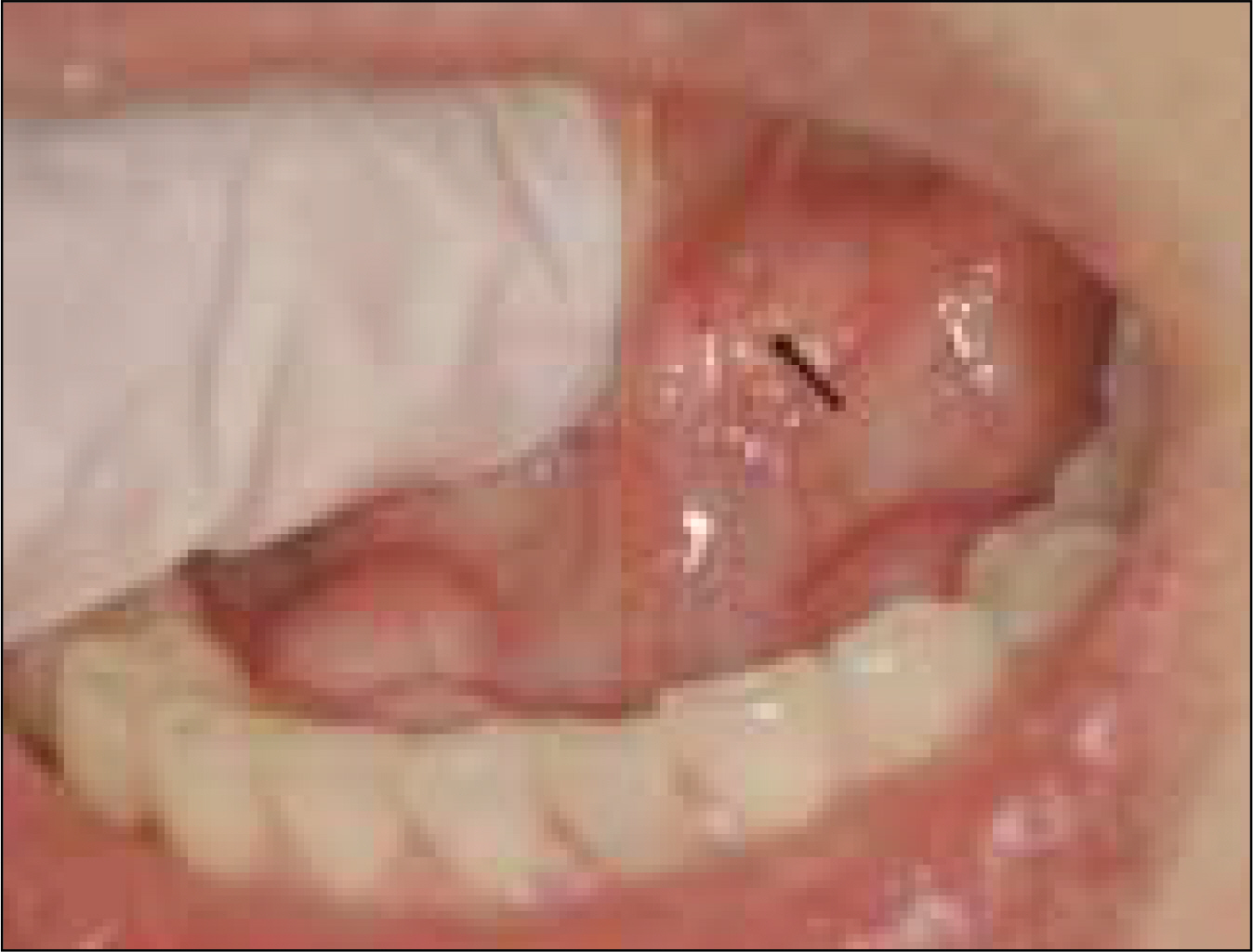
Fig. 2. Pre-operative intraoral view. A firm material was palpated. (arrow)
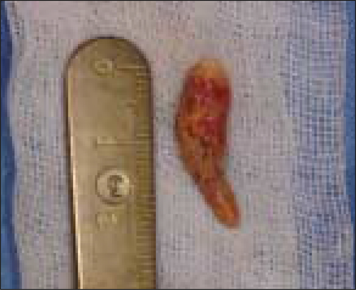
Fig. 3. Macroscopic view of giant sialolith. It measured to be approximately 22 mm in length, 5.5 mm in wide.
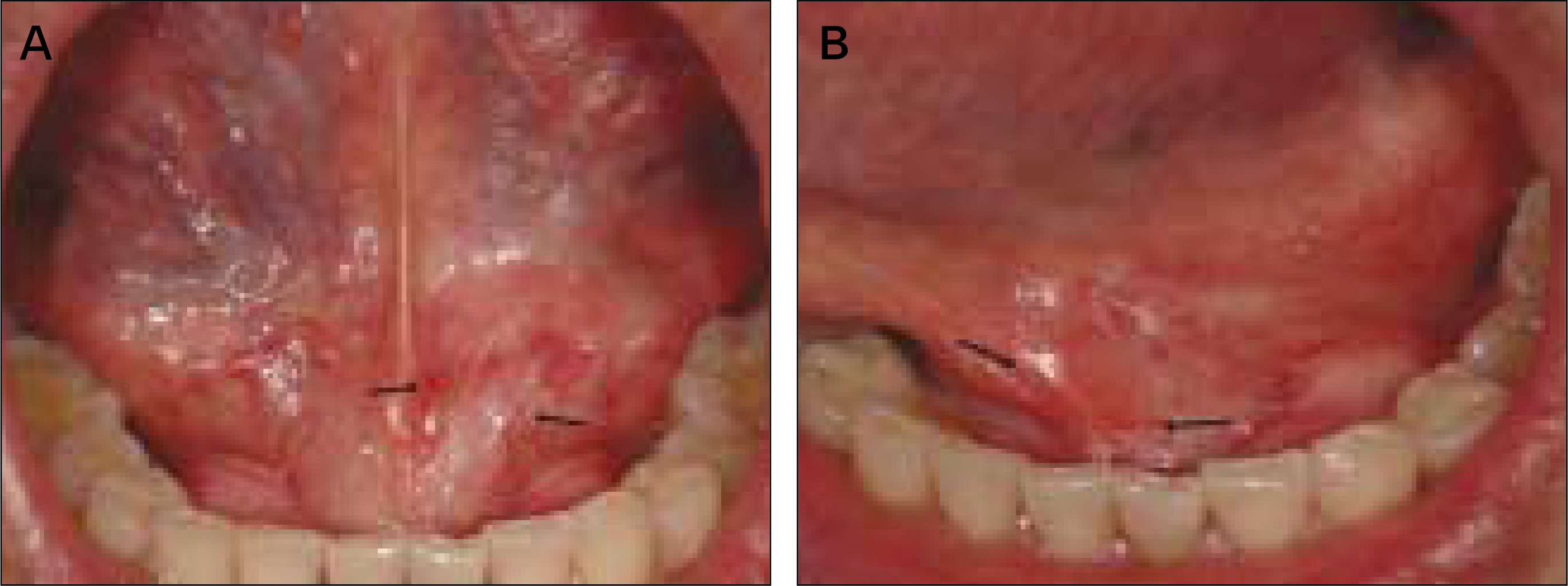
Fig. 4. A. Post-operative intraoral anterior view (8 month), B. Left side view. (8 month) Scar was formed on operation site, but saliva was discharged to Wharton's duct & operation site. Patient had no clinical symptom. Salivary flow was normal. (Right arrow: Wharton's duct orifice, Left arrow: operation site fistula)
Cited by 1 articles
-
Removal of submandibular calculi by surgical method and hydraulic power with curved needle: a case report
Seong-Ho Cho, Ji-Deuk Han, Jung-Han Kim, Shi-Hyun Lee, Ji-Bong Jo, Chul-Hoon Kim, Bok-Joo Kim
J Korean Assoc Oral Maxillofac Surg. 2017;43(3):182-185. doi: 10.5125/jkaoms.2017.43.3.182.
Reference
-
References
1. Ord RA, Pazoki AE. Salivary Gland Disease and Tumors. Miloro M, editor. Peterson's principles of oral and maxillofacial surgery. 2nd ed.London: BC Decker Inc.;2004. p. 671–7.2. Lustmann J, Regev E, Melamed Y. Sialolithiasis. A survey on 245 patients and a review of the literature. Int J Oral Maxillofac Surg. 1990; 19:135–8.3. Bodner L. Giant salivary gland calculi: diagnostic imaging and surgical management. Oral Surg Oral Med Oral Pathol Oral Radiol Endod. 2002; 94:320–3.
Article4. Rai M, Burman R. Giant submandibular sialolith of remarkable size in the comma area of Wharton's duct: a case report. J Oral Maxillofac Surg. 2009; 67:1329–32.
Article5. Baurmash HD. Submandibular salivary stones: current management modalities. J Oral Maxillofac Surg. 2004; 62:369–78.
Article6. Combes J, Karavidas K, McGurk M. Intraoral removal of proximal submandibular stones – an alternative to sialadenectomy? Int J Oral Maxillofac Surg. 2009; 38:813–6.
Article7. Nahlieli O, Shacham R, Zagury A, Bar T, Yoffe B. The ductal stretching technique: an endoscopic-assisted technique for removal of submandibular stones. Laryngoscope. 2007; 117:1031–5.
Article8. Ardekian L, Klain H, Peled M. Obstructive sialadenitis of submandibular gland due to foreign body successfully treated by sialoendoscopic intervention. J Oral Maxillofac Surg. 2009; 67:1337–9.
Article9. Liu DG, Zhang ZY, Zhang Y, Zhang L, Yu GY. Diagnosis and management of sialolithiasis with a semirigid endoscope. Oral Surg Oral Med Oral Pathol Oral Radiol Endod. 2009; 108:9–14.
Article10. Escudier MP, Brown JE, Drage NA, McGurk M. Extracorporeal shockwave lithotripsy in the management of salivary calculi. Br J Surg. 2003; 90:482–5.
Article11. Siddiqui SJ. Sialolithiasis: an unusually large submandibular salivary stone. Br Dent J. 2002; 193:89–91.
Article12. Chung IK, Kim JR, Kim UK, Shin SH, Kim YD, Byun JH, et al. A clinical study of submandibular gland excision. J Kor Oral Maxillofac Surg. 2004; 30:545–550.13. Ledesma-Montes C, Garce ′ s-Ortl′ z Salcido-Garcl′ a JF, Herna ′ – ndez-Flores F, Herna ′ ndez-Guerrero JC. Giant sialolith: case report and review of the literature. J Oral Maxillofac Surg. 2007; 65:128–30.
Article14. Boffano P, Gallesio C. Surgical treatment of a giant sialolith of the Wharton duct. J Craniofac Surg. 2010; 21:134–5.
Article15. Soares EC, Costa FW, Pessoa RM, Bezerra TP. Giant salivary calculus of the submandibular gland. Otolaryngol Head Neck Surg. 2009; 140:128–9.
Article16. Zakaria MA. Giant calculi of the submandibular salivary gland. Br J Oral Surg. 1981; 19:230–2.
Article17. Iqbal SM, Murthy JG, Sharma N. Giant parotid calculus-an unusual presentation. J Laryngol Otol. 1992; 106:446–7.
Article18. Kesse WK, Shehab ZP, Courteney-Harris R. A megalith of the parotid salivary gland. J Laryngol Otol. 1998; 112:784–5.
Article
- Full Text Links
-
- Actions
-
Cited
- CITED
-
- Close
- Share
-
- Similar articles
-
- A Case of Unilateral Absence of the Submandibular Gland Secondary to Sialolithiasis
- A giant sialolith in a wharton's duct: A case report
- Sialolithiasis Mimicking Metastatic Thyroid Cancer
- Recurrent Sialolithiasis on Remnant Wharton's Duct Following Submandibular Gland Resection
- A case report of the sialolithiasis on the submandibular gland
