J Korean Assoc Oral Maxillofac Surg.
2013 Feb;39(1):35-40. 10.5125/jkaoms.2013.39.1.35.
Giant osteochondroma of the parapharyngeal space: a case report
- Affiliations
-
- 1Department of Oral and Maxillofacial Surgery, School of Dentistry, Dankook University, Cheonan, Korea. kimchoms@dankook.ac.kr
- KMID: 2189563
- DOI: http://doi.org/10.5125/jkaoms.2013.39.1.35
Abstract
- Osteochondroma is a common benign tumor of the axial skeleton, especially in the distal metaphysis of the femur and the proximal metaphysis of the tibia, that can occur on the facial skeleton (albeit rarely). Osteochondroma is differentiated from chondroma, osteochondromatosis and osteoma. Osteochondroma shows an irregular radiopaque lesion and chondromatic area surrounded by the osteoma. When it develops in the long bone, it has a marked tendency to occur at 10 to 20 years of age and ceases with the end of pubertal growth. However, when it develops in the mandibular condyle, it is prevalent in the third decade and continuous to develop. Tumors that develop in the long bone have a predilection for men, but tumors in the mandible have a predilection for women. In osteochondroma of the mandibular condyle, clinical features presented include occlusal changes, facial asymmetry, headaches, pain and joint noise on the temporomandibular joint, mouth opening limitations, and jaw deviation at the involved site. The first choice of treatment for the massive osteochondroma is surgical removal. A 70-year-old female patient with an osteochondroma on her right mandibular condyle visited our clinic. We surgically removed the mass with favorable results. It is presented here along with a review of literature on osteochondroma.
Keyword
MeSH Terms
Figure
-
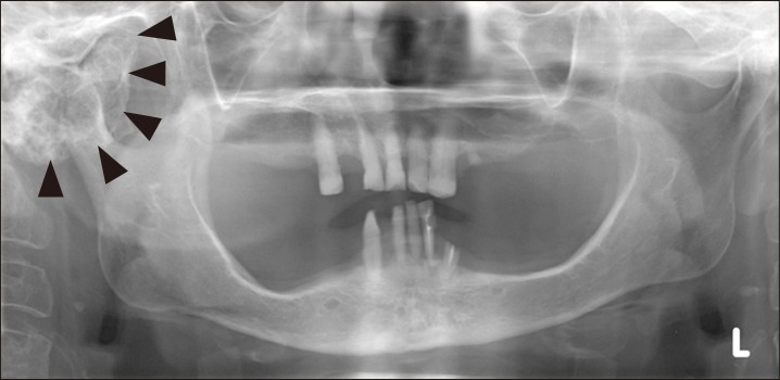
Fig. 1 Initial panoramic view shows round radiopaque mass on the right mandibular condyle with jaw deviation (arowheads).
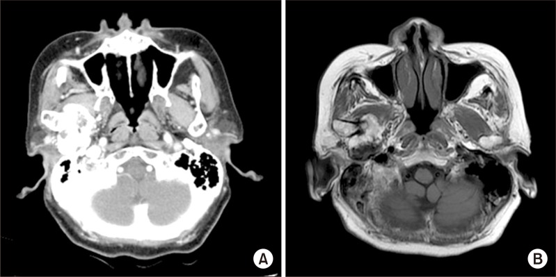
Fig. 2 Preoperative computed tomography (A), magnetic resonance imaging (B). Axial view shows mass invaded right parapharyngeal space.
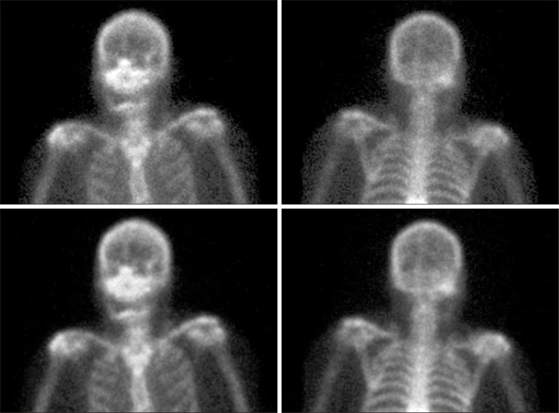
Fig. 3 99mTc bone scan. There is a focal hot spot in the right mandibular condyle
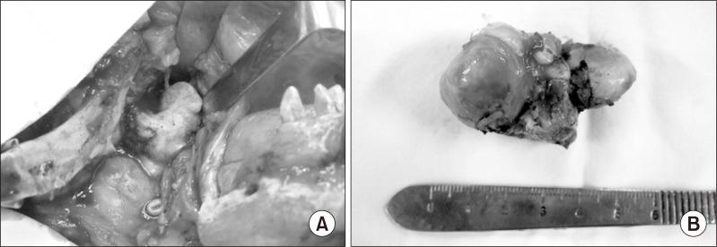
Fig. 4 Intraoperative finding. A. Exposure hard tumor mass in skull base via lip-split incision. B. 5×3×3 cm size bony mass was removed.
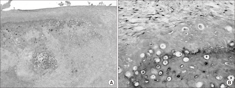
Fig. 5 A. Photomicrograph shows cartilaginous cap and endochondral ossification. B. Magnification of the area of endochondral ossification. (H&E staining, A: ×40, B: ×100)

Fig. 6 Postoperative computed tomography. Axial view.
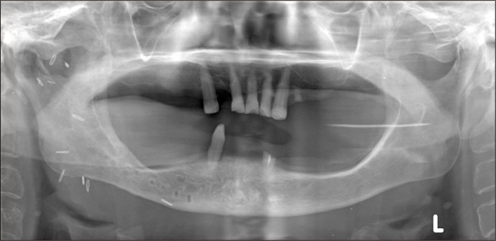
Fig. 7 Postoperative panorama view.
Reference
-
1. Meyer RA. Osteochondroma of coronoid process of mandible: report of case. J Oral Surg. 1972; 30:297–300. PMID: 4501965.2. Huh HY, Jo SG, Jin WJ, Shin HK, Kim OW. Cases report of surgical correction of unilateral osteochondroma. J Korean Acad Oral Surg. 1987; 13:57–66.3. Landini G, Kitano M, Urago A, Sugihara K, Yamashita S. Chondroma and osteochondroma of the tongue. Oral Surg Oral Med Oral Pathol. 1989; 68:206–209. PMID: 2780021.
Article4. Traub DJ, Marco WP, Eisenberg E, Barrows G. Osteochondroma of the maxillary sinus: report of a case. J Oral Maxillofac Surg. 1990; 48:752–755. PMID: 2358955.
Article5. Pool JW, Tilson HB, Thornton WE, Steed DL. Osteochondroma of the zygomatic arch: report of case. J Oral Surg. 1979; 37:673–675. PMID: 288891.6. Barnes L. Surgical pathology of the head and neck. 1985. New York: Dekker.7. Lichtenstein L. Classification of primary tumors of bone. Cancer. 1951; 4:335–341. PMID: 14821927.
Article8. Marx RE, Stern D. Oral and maxillofacial pathology : a rationale for diagnosis and treatment. 2003. Chicago: Quintessence;p. 772.9. Peroz I, Scholman HJ, Hell B. Osteochondroma of the mandibular condyle: a case report. Int J Oral Maxillofac Surg. 2002; 31:455–456. PMID: 12361086.
Article10. Gaines RE Jr, Lee MB, Crocker DJ. Osteochondroma of the mandibular condyle: case report and review of the literature. J Oral Maxillofac Surg. 1992; 50:899–903. PMID: 1634981.
Article11. Langenskiöld A. The development of multiple cartilaginous exostoses. Acta Orthop Scandinav. 1967; 38:259–266.
Article12. Kim MS, Lee MH, Jang CS, Kim CH. A case report of osteochondroma on mandibular condyle. J Korean Assoc Maxillofac Plast Reconstr Surg. 1996; 18:298–307.13. Allan JH, Scott J. Osteochondroma of the mandible. Oral Surg Oral Med Oral Pathol. 1974; 37:556–565. PMID: 4521947.
Article14. Kaneda T, Torii S, Yamashita T, Inoue N, Shimizu K. Giant osteochondroma of the mandibular condyle. J Oral Maxillofac Surg. 1982; 40:818–821. PMID: 6958845.
Article15. Huvos AG. Bone tumors, diagnosis, treatment, and prognosis. 1979. Philadelphia: Saunders;p. 139–160.16. Forssell H, Happonen RP, Forssell K, Virolainen E. Osteochondroma of the mandibular condyle. Report of a case and review of the literature. Br J Oral Maxillofac Surg. 1985; 23:183–189. PMID: 3159417.
Article17. Vezeau PJ, Fridrich KL, Vincent SD. Osteochondroma of the mandibular condyle: literature review and report of two atypical cases. J Oral Maxillofac Surg. 1995; 53:954–963. PMID: 7629631.18. Henry CH, Granite EL, Rafetto LK. Osteochondroma of the mandibular condyle: report of a case and review of the literature. J Oral Maxillofac Surg. 1992; 50:1102–1108. PMID: 1527664.
Article19. Marks RB, Carlton DM Jr, Carr RF. Osteochondroma of the mandibular condyle. Report of a case with 10-year follow-up. Oral Surg Oral Med Oral Pathol. 1984; 58:30–32. PMID: 6589574.
- Full Text Links
-
- Actions
-
Cited
- CITED
-
- Close
- Share
-
- Similar articles
-
- Giant Intra-articular Osteochondroma of the Knee: A Case Report
- Giant Osteochondroma from the Rib: A report of One Case
- The Development of a Giant Extraskeletal Osteochondroma in the Masticatory Space of the Mandible
- Osteochondroma of the Cervical Spine: A Case Report
- Osteochondroma of the Talus: A Report of Two Cases
