An alternative treatment option for a bony defect from large odontoma using recycled demineralization at chairside
- Affiliations
-
- 1Department of Oral and Maxillofacial Surgery, Dankook University Jukjeon Dental Hospital, Yongin, Korea. eskimos@dankook.ac.kr
- 2Department of Oral and Maxillofacial Surgery, Chungbuk National University College of Medicine, Cheongju, Korea.
- 3Department of Prosthodontics, Ewha Womans University School of Medicine, Seoul, Korea.
- KMID: 2189476
- DOI: http://doi.org/10.5125/jkaoms.2015.41.2.109
Abstract
- Odontoma is the most common odontogenic benign tumor, and the treatment of choice is generally surgical removal. After excision, bone grafts may be necessary depending on the need for further treatment, or the size and location of the odontoma. Although the osteogenic capacity of a demineralized tooth was verified as early as 1967 by Urist and many other investigators, the cumbersome procedure, including a long demineralization time, may be less than comfortable for clinicians. A modified ultrasonic technology, with periodic negative pressure and temperature control, facilitated rapid and aseptic preparation of demineralized teeth for bone grafts. This approach reduces the demineralization time dramatically (< or =80 minutes), so that the graft material can be prepared chairside on the same day as the extraction. The purpose of this article is to describe two cases of large compound odonotomas used as graft material prepared chairside for enucleation-induced bony defects. These two clinical cases showed favorable wound healing without complications, and good bony support for future dental implants or orthodontic treatment. Finally, this report will suggest the possibility of recycling the benign pathologic hard tissue as an alternative treatment option for conventional bone grafts in clinics.
Keyword
MeSH Terms
Figure
-

Fig. 1 Preoperative panoramic radiopraph (A) and computed tomography (CT) scans (B, C) show compound odontoma associated with failure of permanent canine eruption. A. A odontoma is circumscribed by a radiolucent halo underneath an impacted canine (arrows). B. CT scans in axial section show that the alveolar bone was bulged to labial side slightly (arrows). C. CT scans in coronal section show an odontoma with its superior limits invading the floor of the nasal cavity and its posterior limits in the hard palate, and impacted the right maxillary canine above the lesion (arrows).
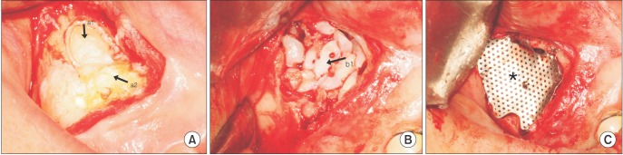
Fig. 2 Intraoral photos during operation. After opening the alveolar bony window (A), demineralized odontoma and tooth block were grafted followed by surgical removal (B), and titanium mesh filled with bone graft and secured into position with screws in the area of the bony defect (C). (a1: an impacted maxillary canine, a2: an odontoma, b1: demineralized odontoma and tooth block, asterisk: titanium mesh)
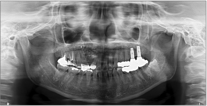
Fig. 3 Postoperative panoramic radiograph showing the titanium mesh in position with demineralized tooth graft materials.
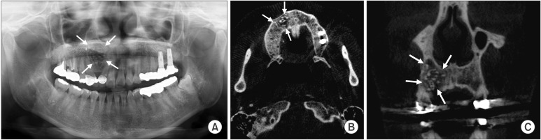
Fig. 4 Postoperative panoramic radiograph (A) and computed tomography scans in coronal (B), axial (C) section at 6-months follow-up shows favorable new bone formation and subsequent resorption of graft materials (arrows).
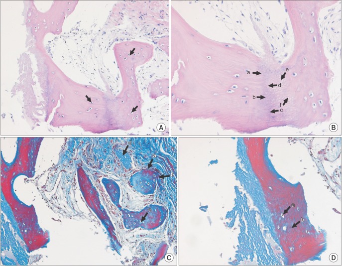
Fig. 5 The section in 6-months was stained with H&E staining (A: ×200, B: ×400) and Masson's trichorme staining (C: ×200, D: 400). Osteocytic embedding in graft tooth material (A, arrows), intimate fusion between new bone and graft tooth material (B, a-f) is observed. Osteocytic embedding in graft material with new bone were shown. Embedded osteocyte (C, c; D, a), newly formed bone (C, a,b,d; D, b) were also confirmed in the specimens.
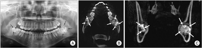
Fig. 6 Preoperative panoramic radiopgraph (A) and computed tomography scans (B, C) show a compound odontoma with impacted 3rd molar, contacting above the bony canal of inferior alveolar nerve (left, 4 arrows). Right impacted 3rd molar (right, 3 arrows) was also extracted for the orthodontic treatment.

Fig. 7 Postoperative panoramic radiograph at postoperative 1-day (A), and 1-year (B), computed tomography scans (C, D) at 1-year follow-up. Radiographs at 6-months (B-D) showed favorable new bone formation, regularity of alveolar ridge, and higher bony density of graft site. Patient could start orthodontic treatment after 1 month of mass excision. Left: 4 arrows, right: 3 arrows.
Cited by 3 articles
-
Histomorphometric study of rabbit's maxillary sinus augmentation with various graft materials
Dong-Seok Sohn, Yong-Suk Moon
Anat Cell Biol. 2018;51(Suppl 1):S1-S12. doi: 10.5115/acb.2018.51.S1.S1.Giant complex odontoma in the posterior mandible: A case report and literature review
Jong Chan Park, Ji Ho Yang, Sung Youn Jo, Bong Chul Kim, Jun Lee, Wan Lee
Imaging Sci Dent. 2018;48(4):289-293. doi: 10.5624/isd.2018.48.4.289.Comparison of immunohistochemical analysis on sinus augmentation using demineralized tooth graft and bovine bone
Dong-Seok Sohn, Ji-Rak Kim, Hyung-Gyun Kim, Hyun-Suk Choi, Yong-Suk Moon
J Korean Assoc Oral Maxillofac Surg. 2021;47(4):269-278. doi: 10.5125/jkaoms.2021.47.4.269.
Reference
-
1. Budnick SD. Compound and complex odontomas. Oral Surg Oral Med Oral Pathol. 1976; 42:501–506. PMID: 1067549.
Article2. Neville B, Damm DD, Allen CM, Bouquot J. Oral and maxillofacial pathology. Philadelphia: WB Saunders;2007.3. Owens BM, Schuman NJ, Mincer HH, Turner JE, Oliver FM. Dental odontomas: a retrospective study of 104 cases. J Clin Pediatr Dent. 1997; 21:261–264. PMID: 9484137.4. Mehra P, Singh H. Complex composite odontoma associated with impacted tooth: a case report. N Y State Dent J. 2007; 73:38–40. PMID: 17472184.5. Louis PJ, Gutta R, Said-Al-Naief N, Bartolucci AA. Reconstruction of the maxilla and mandible with particulate bone graft and titanium mesh for implant placement. J Oral Maxillofac Surg. 2008; 66:235–245. PMID: 18201602.
Article6. Yeomans JD, Urist MR. Bone induction by decalcified dentine implanted into oral, osseous and muscle tissues. Arch Oral Biol. 1967; 12:999–1008. PMID: 4226721.
Article7. Kim YK, Kim SG, Byeon JH, Lee HJ, Um IU, Lim SC, et al. Development of a novel bone grafting material using autogenous teeth. Oral Surg Oral Med Oral Pathol Oral Radiol Endod. 2010; 109:496–503. PMID: 20060336.
Article8. Murata M, Akazawa T, Takahata M, Ito M, Tazaki J, Hino J, et al. Bone induction of human tooth and bone crushed by newly developed automatic mill. J Ceramic Soc Jpn. 2010; 118:434–437.
Article9. Kim YK, Kim SG, Yun PY, Yeo IS, Jin SC, Oh JS, et al. Autogenous teeth used for bone grafting: a comparison with traditional grafting materials. Oral Surg Oral Med Oral Pathol Oral Radiol. 2014; 117:e39–e45. PMID: 22939321.
Article10. Nanci A. Ten Cate's oral histology: development, structure and function. 8th ed. Mosby Elsevier;2008. p. 108–192.11. Butler WT, Mikulski A, Urist MR, Bridges G, Uyeno S. Noncollagenous proteins of a rat dentin matrix possessing bone morphogenetic activity. J Dent Res. 1977; 56:228–232. PMID: 265954.
Article12. Liu Y, Luo D, Liu S, Fu Y, Kou X, Wang X, et al. Effect of nanostructure of mineralized collagen scaffolds on their physical properties and osteogenic potential. J Biomed Nanotechnol. 2014; 10:1049–1060. PMID: 24749399.
Article13. Beniash E. Biominerals--hierarchical nanocomposites: the example of bone. Wiley Interdiscip Rev Nanomed Nanobiotechnol. 2011; 3:47–69. PMID: 20827739.
Article14. Behrend O, Schubert H. Influence of hydrostatic pressure and gas content on continuous ultrasound emulsification. Ultrason Sonochem. 2001; 8:271–276. PMID: 11441610.
Article15. Fang QG, Shi S, Sun CF. Odontogenic lesions in pediatric patients. J Craniofac Surg. 2014; 25:e248–e251. PMID: 24785745.
Article16. Fasolis M, Boffano P, Ramieri G. Morbidity associated with anterior iliac crest bone graft. Oral Surg Oral Med Oral Pathol Oral Radiol. 2012; 114:586–591. PMID: 22901642.
Article17. Atiya BK, Shanmuhasuntharam P, Huat S, Abdulrazzak S, Oon H. Liquid nitrogen-treated autogenous dentin as bone substitute: an experimental study in a rabbit model. Int J Oral Maxillofac Implants. 2014; 29:e165–e170. PMID: 24683581.
Article18. Rezende ML, Consolaro A, Sant'Ana AC, Damante CA, Greghi SL, Passanezi E. Demineralization of the contacting surfaces in autologous onlay bone grafts improves bone formation and bone consolidation. J Periodontol. 2014; 85:e121–e129. PMID: 24171500.
Article19. Suzuki S, Haruyama N, Nishimura F, Kulkarni AB. Dentin sialophosphoprotein and dentin matrix protein-1: two highly phosphorylated proteins in mineralized tissues. Arch Oral Biol. 2012; 57:1165–1175. PMID: 22534175.
Article20. Huang YF, Meek KM, Wang LQ, Wang DJ. Effects of prior freezing or drying on the swelling behaviour of the bovine cornea. Chin Med J (Engl). 2009; 122:212–218. PMID: 19187649.
- Full Text Links
-
- Actions
-
Cited
- CITED
-
- Close
- Share
-
- Similar articles
-
- Discussion: An alternative treatment option for a bony defect from large odontoma using recycled demineralization at chairside
- Incidentally detected odontoma within a dentigerous cyst
- Unusually large erupted complex odontoma: A rare case report
- Ameloblastic fibro-odontoma: a case report
- Ameloblastic fibro-odontoma in the mandible: a case report
