Validity of the vertical tube-shift method in determining the relationship between the mandibular third molar roots and the inferior alveolar nerve canal
- Affiliations
-
- 1Department of Oral Medicine and Radiology, The Oxford Dental College, Bangalore, India. dr.anjanaarora2909@gmail.com
- KMID: 2189465
- DOI: http://doi.org/10.5125/jkaoms.2015.41.2.66
Abstract
OBJECTIVES
To assess the validity of the vertical tube-shift method using intraoral periapical radiography (IOPAR) for determining the relationship between the mandibular third molar roots and the inferior alveolar nerve (IAN) canal in comparison with cone-beam computed tomography (CBCT).
MATERIALS AND METHODS
Fifty impacted mandibular third molars were analyzed using the IOPAR vertical tube-shift method and CBCT. The relationship of the IAN canal to the impacted mandibular third molar was recorded as buccal, lingual or in line with the apex and was compared with CBCT findings. The sensitivity, specificity, positive predictive value (PPV), and negative predictive value (NPV) of the vertical tube-shift method in depicting the relationship (buccal/lingual/in line with the apex) of the IAN canal to the third molar root apex was calculated.
RESULTS
The sensitivity and specificity PPV and NPV of the IOPAR vertical tube-shift technique was found to be highest for a lingual relationship (100%) followed by buccal (94.4%, 92.3%, 97.1%, and 85.7%) and in line with the apex relationship (88.9%, 95.0%, 80.0%, and 97.4%) of the IAN canal with the third molar root apex, respectively. A statistically significant association was observed between the IOPAR vertical tube-shift method and the CBCT with a P-value <0.01.
CONCLUSION
The vertical tube-shift method can be used as an effective diagnostic tool in assessing the relationship of the IAN canal to the third molar root apex with high sensitivity, specificity, PPV, and NPV.
Keyword
MeSH Terms
Figure
-
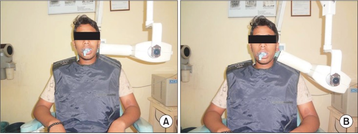
Fig. 1 Intraoral periapical radiography exposure was made by directing the position-indicating device at 0° (A) and -20° (B) vertical angulations. Informed consent was obtained from patient willing to participate in the present study.
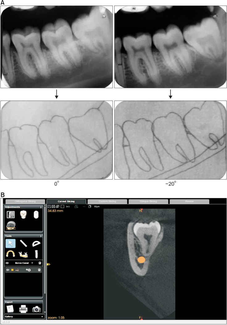
Fig. 2 A. Intraoral periapical radiography at 0°, -20° by vertical tube-shift method and their tracing showing the inferior alveolar nerve (IAN) canal moving upwards predicting the canal as buccal to the third molar root apex. B. Cone-beam computed tomography of the same patient as in Fig. 2A, showing the IAN canal buccal to the third molar root apex.
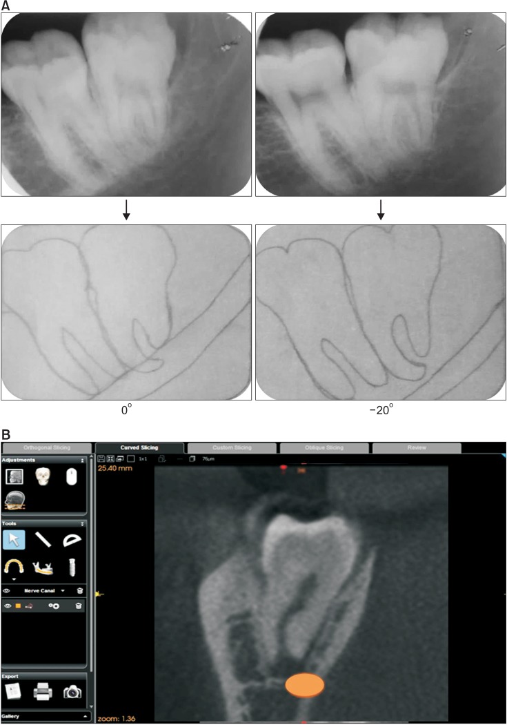
Fig. 3 A. Intraoral periapical radiography at 0°, -20° by vertical tube-shift method and their tracing showing inferior alveolar nerve (IAN) canal moving downwards predicting canal lingual to the third molar root apex. B. Cone-beam computed tomography of the same patient as in Fig. 3A, showing the IAN canal lingual to the third molar root apex.
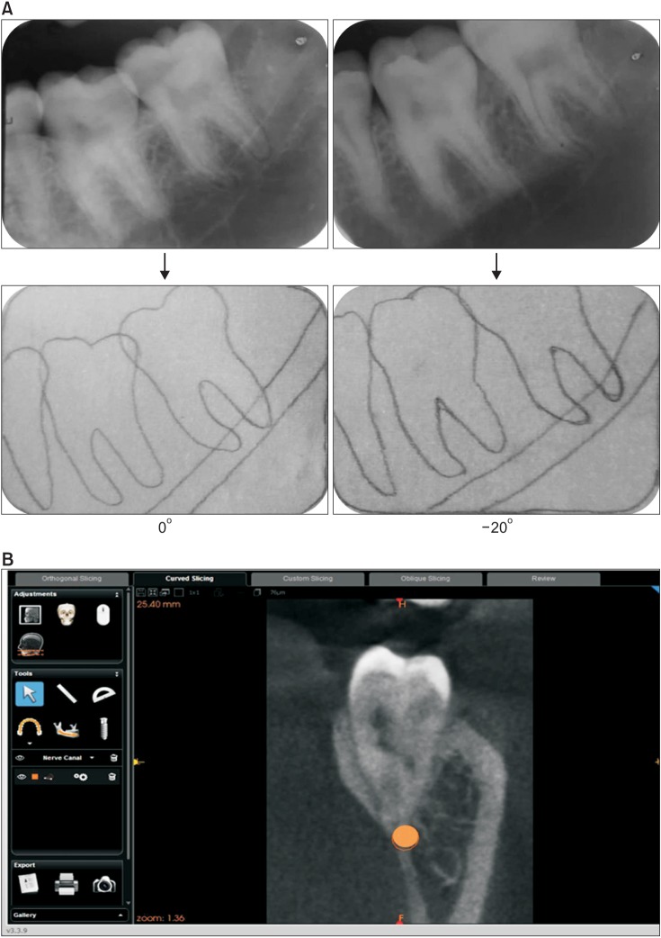
Fig. 4 A. Intraoral periapical radiography at 0°, -20° by vertical tube-shift method and their tracing showing no change in the inferior alveolar nerve (IAN) canal position relative to the third molar root apex. B. Cone-beam computed tomography of the same patient as in Fig. 4A showing no change in the IAN canal position relative to the third molar root apex.
Cited by 1 articles
-
Preoperative imaging of the inferior alveolar nerve canal by cone-beam computed tomography and 1-year neurosensory recovery following mandibular setback through bilateral sagittal split ramus osteotomy: a randomized clinical trial
Ali Hassani, Vahid Rakhshan, Mohammad Hassani, Hamidreza Mahaseni Aghdam
J Korean Assoc Oral Maxillofac Surg. 2020;46(1):41-48. doi: 10.5125/jkaoms.2020.46.1.41.
Reference
-
1. Neves FS, Souza TC, Almeida SM, Haiter-Neto F, Freitas DQ, Bóscolo FN. Correlation of panoramic radiography and cone beam CT findings in the assessment of the relationship between impacted mandibular third molars and the mandibular canal. Dentomaxillofac Radiol. 2012; 41:553–557. PMID: 22282507.
Article2. Szalma J, Lempel E, Jeges S, Szabó G, Olasz L. The prognostic value of panoramic radiography of inferior alveolar nerve damage after mandibular third molar removal: retrospective study of 400 cases. Oral Surg Oral Med Oral Pathol Oral Radiol Endod. 2010; 109:294–302. PMID: 19846324.
Article3. Neugebauer J, Shirani R, Mischkowski RA, Ritter L, Scheer M, Keeve E, et al. Comparison of cone-beam volumetric imaging and combined plain radiographs for localization of the mandibular canal before removal of impacted lower third molars. Oral Surg Oral Med Oral Pathol Oral Radiol Endod. 2008; 105:633–642. PMID: 18299225.
Article4. Tantanapornkul W, Okochi K, Bhakdinaronk A, Ohbayashi N, Kurabayashi T. Correlation of darkening of impacted mandibular third molar root on digital panoramic images with cone beam computed tomography findings. Dentomaxillofac Radiol. 2009; 38:11–16. PMID: 19114418.
Article5. Marciani RD. Third molar removal: an overview of indications, imaging, evaluation, and assessment of risk. Oral Maxillofac Surg Clin North Am. 2007; 19:1–13. PMID: 18088860.
Article6. Kositbowornchai S, Densiri-aksorn W, Piumthanaroj P. Ability of two radiographic methods to identify the closeness between the mandibular third molar root and the inferior alveolar canal: a pilot study. Dentomaxillofac Radiol. 2010; 39:79–84. PMID: 20100918.
Article7. Akçiçek G, Uysal S, Avcu N, Kansu Ö. Comparison of different imaging techniques for the evaluation of proximity between molars and the mandibular canal. Clin Dent Res. 2012; 36:2–7.8. Jaju PP. Localization of mandibular canal by buccal object rule. Oral Surg Oral Med Oral Pathol Oral Radiol Endod. 2010; 109:799. PMID: 20451840.
Article9. Morant RD, Eleazer PD, Scheetz JP, Farman AG. Array-projection geometry and depth discrimination with Tuned-Aperture Computed Tomography for assessing the relationship between tooth roots and the inferior alveolar canal. Oral Surg Oral Med Oral Pathol Oral Radiol Endod. 2001; 91:252–259. PMID: 11174606.
Article10. Richards AG. Roentgenographic localization of the mandibular canal. J Oral Surg (Chic). 1952; 10:325–329. PMID: 12991124.11. Tantanapornkul W, Okouchi K, Fujiwara Y, Yamashiro M, Maruoka Y, Ohbayashi N, et al. A comparative study of cone-beam computed tomography and conventional panoramic radiography in assessing the topographic relationship between the mandibular canal and impacted third molars. Oral Surg Oral Med Oral Pathol Oral Radiol Endod. 2007; 103:253–259. PMID: 17234544.
Article12. Shahidi S, Zamiri B, Bronoosh P. Comparison of panoramic radiography with cone beam CT in predicting the relationship of the mandibular third molar roots to the alveolar canal. Imaging Sci Dent. 2013; 43:105–109. PMID: 23807934.
Article13. Heurich T, Ziegler C, Steveling H, Wörtche R, Mühling J, Hassfeld S. Digital volume tomography--an extension to the diagnostic procedures available for application before surgical removal of third molars. Mund Kiefer Gesichtschir. 2002; 6:427–432. PMID: 12447656.14. Jasa GR, Vizzotto MB, Silveira PF, Silveira HE, Silveira HL, Correa LR, et al. Buccal-lingual localization of the mandibular canal in relationship with the third molar using the lateral oblique technique. J Oral Maxillofac Radiol. 2014; 2:15–20.
Article15. Miller CS, Nummikoski PV, Barnett DA, Langlais RP. Crosssectional tomography. A diagnostic technique for determining the buccolingual relationship of impacted mandibular third molars and the inferior alveolar neurovascular bundle. Oral Surg Oral Med Oral Pathol. 1990; 70:791–797. PMID: 2263343.
- Full Text Links
-
- Actions
-
Cited
- CITED
-
- Close
- Share
-
- Similar articles
-
- The study of evaluation to relationship between the inferior alveolar nerve and the mandibular third molar by using radiographic image
- Analysis and evaluation of relative positions of mandibular third molar and mandibular canal impacts
- Assessment of the relationship between the mandibular third molar and the mandibular canal using panoramic radiograph and cone beam computed tomography
- Study On The Relationship Of The Inferior Alveolar Nerve Position Between Buccal And Lingual Side Using Ct And Orthpantomogram
- Radiographic evaluation of the course and visibility of the mandibular canal
