Neurilemmoma in the floor of the mouth: a case report
- Affiliations
-
- 1Department of Oral and Maxillofacial Surgery, School of Dentistry, Seoul National University, Seoul, Korea. myoungh@snu.ac.kr
- KMID: 2189420
- DOI: http://doi.org/10.5125/jkaoms.2016.42.1.60
Abstract
- Neurilemmomas are well-encapsulated, benign, slow-growing tumors originating from Schwann cells of the nerve sheath surrounding cranial, peripheral, or autonomic nerves. Intraoral neurilemmomas are relatively rare and have a wide variety of morphologic and radiologic features. This makes differential diagnosis difficult, and only histopathological features can lead to a definitive neurilemmoma diagnosis. In this report, we present the case of a 30-year-old woman whose chief complaint was a solitary, nodular mass on the right floor of the mouth. After computed tomography and magnetic resonance imaging, we performed an incisional biopsy that showed the typical characteristics of a neurilemmoma. The mass was removed completely through an intraoral surgical approach. Despite losing a portion of the lingual nerve, the patient did not complain of any specific discomfort. Wound healing was uneventful and there were no signs or symptoms of recurrence.
Keyword
MeSH Terms
Figure
-

Fig. 1 Preoperative intraoral clinical photograph.
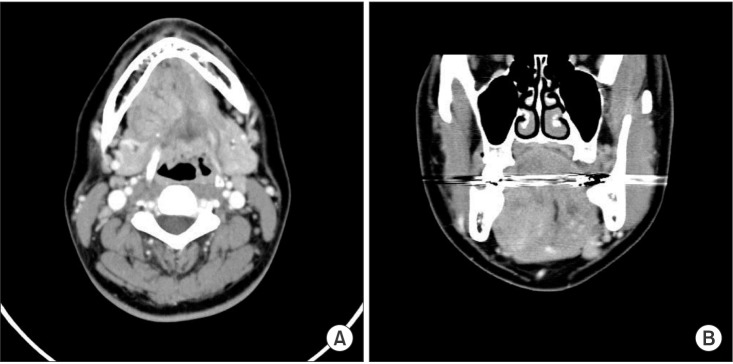
Fig. 2 Computed tomography showing a heterogeneous enhancing lesion. A. Axial view. B. Coronal view. The artifact was seen through all cuts.
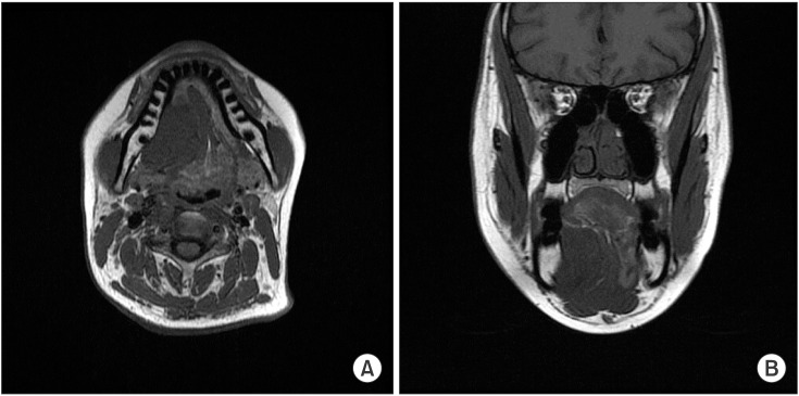
Fig. 3 T1-weighted magnetic resonance imaging showing a well-defined heterogeneous lesion. A. Axial view. B. Coronal view.
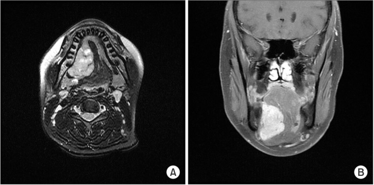
Fig. 4 T2-weighted magnetic resonance imaging showing a well-defined heterogeneous lesion. A. Axial view. B. Coronal view.

Fig. 5 Perioperative clinical photograph. A. The mass was removed through an intraoral approach. B. The mass was well-encapsulated and removed as a whole.
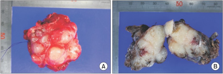
Fig. 6 Postoperative photograph of main mass. A. The mass was well-encapsulated and ovoid in shape. B. Upon dissection of the mass, many exophytic lobules were detected.
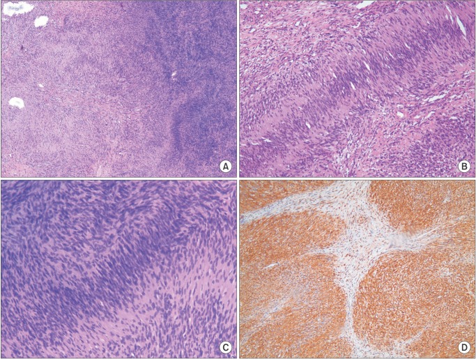
Fig. 7 Microscopic examination of the mass. A. Antoni B pattern with less cellularity, left. Antoni A pattern with well-organized, high cellularity, right (H&E staining, ×40). B. Streaming fascicles of spindle-shaped Schwann cells surround a central acellular, eosinophilic region of Verocay bodies (H&E staining, ×100). C. Magnification of the Antoni A area shows no evidence of cellular dysplasia in spite of high cellularity (H&E staining, ×100). D. Immunohistochemistry shows reactivity to S-100 protein (×100).
Cited by 2 articles
-
Buccal nerve schwannoma mimicking a salivary gland tumor: a rare case report
Jeong-Kui Ku, Dawool Han, Jong-Ki Huh, Jae-Young Kim
J Korean Assoc Oral Maxillofac Surg. 2023;49(3):148-151. doi: 10.5125/jkaoms.2023.49.3.148.A rare histopathological variant of Schwannoma with rosette-like arrangements and epithelioid cells: a case report from a histopathologist’s perspective
Monica Mehendiratta, Vikas Kumar Sant, Manisha Lakhanpal, Keerti Chauhan
J Korean Assoc Oral Maxillofac Surg. 2023;49(4):233-238. doi: 10.5125/jkaoms.2023.49.4.233.
Reference
-
1. Arda HN, Akdogan O, Arda N, Sarikaya Y. An unusual site for an intraoral schwannoma: a case report. Am J Otolaryngol. 2003; 24:348–350. PMID: 13130451.
Article2. Naik SM, Goutham MK, Ravishankara S, Appaji MK. Sublingual Schwannoma: a rare clinical entity reported in a hypothyroid female. Int J Head Neck Surg. 2012; 3:33–39.
Article3. Hsu YC, Hwang CF, Hsu RF, Kuo FY, Chien CY. Schwannoma (neurilemmoma) of the tongue. Acta Otolaryngol. 2006; 126:861–865. PMID: 16846930.
Article4. Tsushima F, Sawai T, Kayamori K, Okada N, Omura K. Schwannoma in the floor of the mouth: a case report and clinicopathological studies of 10 cases in the oral region. J Oral Maxillofac Surg Med Pathol. 2012; 24:175–179.
Article5. Gallo WJ, Moss M, Shapiro DN, Gaul JV. Neurilemoma: review of the literature and report of five cases. J Oral Surg. 1977; 35:235–236. PMID: 264530.6. de Bree R, Westerveld GJ, Smeele LE. Submandibular approach for excision of a large schwannoma in the base of the tongue. Eur Arch Otorhinolaryngol. 2000; 257:283–286. PMID: 10923945.
Article7. Cohen LM, Schwartz AM, Rockoff SD. Benign schwannomas: pathologic basis for CT inhomogeneities. AJR Am J Roentgenol. 1986; 147:141–143. PMID: 3487205.
Article8. La'porte SJ, Juttla JK, Lingam RK. Imaging the floor of the mouth and the sublingual space. Radiographics. 2011; 31:1215–1230. PMID: 21918039.9. AL-Alawi YSM, Kolethekkat AA, Saparamadu PAM, Al Badaai Y. Sublingual gland schwannoma: a rare case at an unusual site. Oman Med J. 2014; 29:DOI: 10.5001/omj.2014.41.
Article10. Rinaldo A, Shaha AR, Pellitteri PK, Bradley PJ, Ferlito A. Management of malignant sublingual salivary gland tumors. Oral Oncol. 2004; 40:2–5. PMID: 14662408.
Article11. Lee YY, Wong KT, King AD, Ahuja AT. Imaging of salivary gland tumours. Eur J Radiol. 2008; 66:419–436. PMID: 18337041.
Article12. Laviv A, Faquin WC, August M. Schwannoma (neurilemmoma) in the floor of the mouth: Presentation of two cases. J Oral Maxillofac Surg Med Pathol. 2015; 27:199–203.
Article13. Neville BW, Damm DD, Allen CM, Bouquot JE. Oral and maxillofacial pathology. 2nd ed. Philadelphia: W.B. Saunders;2002. p. 456–457.14. Enoz M, Suoglu Y, Ilhan R. Lingual schwannoma. J Cancer Res Ther. 2006; 2:76–78. PMID: 17998681.
Article15. Dreher A, Gutmann R, Grevers G. Extracranial schwannoma of the ENT region. Review of the literature with a case report of benign schwannoma of the base of the tongue. HNO. 1997; 45:468–471. PMID: 9324502.16. Daimaru Y, Hashimoto H, Enjoji M. Malignant peripheral nervesheath tumors (malignant schwannomas). An immunohistochemical study of 29 cases. Am J Surg Pathol. 1985; 9:434–444. PMID: 3937453.
