Chondroblastoma in a Distal Phalanx of the Great Toe: A Case Report
- Affiliations
-
- 1Department of Orthopedic Surgery, Korea University Anam Hospital, Seoul, Korea. soonlee@korea.ac.kr
- 2Department of Pathology, Korea University Anam Hospital, Seoul, Korea.
- KMID: 2186480
- DOI: http://doi.org/10.4055/jkoa.2007.42.5.692
Abstract
- A chondroblastoma is a relatively rare benign bone tumor that is typically encountered in the epiphysis. The authors describe a case of a chondroblastoma arising in the great toe. An 82-year-old woman presented with pain over her left great toe with a 1 year duration. The radiographs showed an expansile osteolytic lesion with cortical thinning and coarse trabeculation that replaced the distal phalanx of the left great toe. An incisional biopsy and curettage were performed. Histologically, the tumor consisted of uniform, round to polygonal cells with clear to slightly eosinophilic cytoplasm and round to ovoid nuclei, mimicking chondroblasts, intermingled with osteoclast-like giant cells.
Keyword
MeSH Terms
Figure
-

Fig. 1 Radiographs and 3-dimensional computed tomography images. (A) Radiographs show an ostelytic lesion and course trabeculation in the distal pharynx of the left great toe. (B) 3 dimensional computed tomography shows an expansile osteolytic lesion with cortical thinning out and coarse trabeculation replacing the distal phalanx of the left great toe.
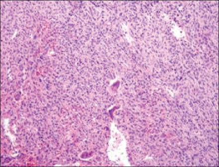
Fig. 2 Curettage specimen shows a uniform to polygonal mononuclear cells with well-defined cytoplasmic borders and slightly eosinophilic cytoplasm intermingled with scattered osteoclast-like giant cells (hematoxylin and eosin, ×200).
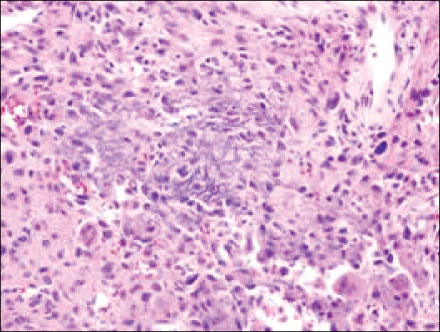
Fig. 3 High-power photomicrograph of the biopsy specimen shows fine calcification around the chondroblasts also known as "chicken-wire calcification" (hematoxylin and eosin, ×400).
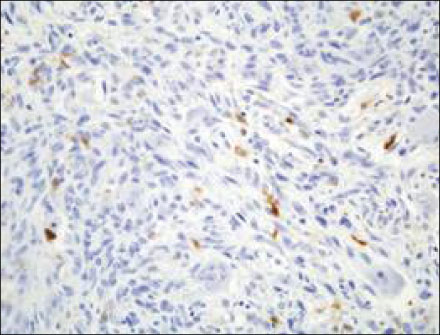
Fig. 4 Some tumor cells tested positive for the S-100 protein.
Reference
-
1. Brien EW, Mirra JM, Ippolito V. Chondroblastoma arising from a nonepiphyseal site. Skeletal Radiol. 1995. 24:220–222.
Article2. Codman EA. The classic: epiphyseal chondromatous giant cell tumors of the upper end of the humerus. Surg Gynecol Obstet. 1931;52:543-548. Clin Orthop Relat Res. 2006. 09. 450:12–16.3. Davila JA, Amrami KK, Sundaram M, Adkins MC, Unni KK. Chondroblastoma of the hands and feet. Skeletal Radiol. 2004. 33:582–587.
Article4. Jaffe HL, Lichtenstein L. Benign chondroblastoma of bone: a reinterpretation of the so-called calcifying or chondromatous giant cell tumor. Am J Pathol. 1942. 18:969–991.5. Castanedo-Cazares JP, Lepe V, Moncada B. Subungual chondroblastoma in a 9-year-old girl. Pediatr Dermatol. 2004. 21:452–453.
Article6. Kang LC, Chamyan G, Barness-Gilbert E. Pathology teach and tell: chondroblastoma. Pediatr Pathol Mol Med. 2002. 21:71–74.7. Kilpatrick SE, Parisien M, Bridge JA. Fletcher CDM, Unni KK, Mertens F, editors. Chondroblastoma. World Health Organization classification of tumours. Pathology and genetics of tumours of soft tissue and bone. 2002. Lyon: IARC Press;241–242.8. Levine GD, Bensch KG. Chondroblastoma--the nature of the basic cell. A study by means of histochemistry, tissue culture, electron microscopy, and autoradiography. Cancer. 1972. 29:1546–1562.9. Romeo S, Bovée JV, Jadnanansing NA, Taminiau AH, Hogendoorn PC. Expression of cartilage growth plate signalling molecules in chondroblastoma. J Pathol. 2004. 202:113–120.
Article10. Welsh RA, Meyer AT. A histogenetic study of chondroblastoma. Cancer. 1964. 17:578–589.
Article
- Full Text Links
-
- Actions
-
Cited
- CITED
-
- Close
- Share
-
- Similar articles
-
- Homodigital Reverse Pedicle Island Flap for Reconstruction of the Great Toe: A Case Report
- Treatment for the Stress Fracture of the Proximal Phalanx of the Great Toe in a Basketball Player with Hallux Valgus (A Case Report)
- Chondroblastoma in Hand: A Case Report
- Fracture Dislocation of the interphalangeal Joint of Great Toe: Report of Three Cases
- A Clinical Observation for the Fracture of Proximal Phalanx of the Great Toe(So Called Safety shoes-Fracture)
