Compressive-Extension Stage 4 Injury in Cervical Spine is Theoretical Stage?: Case Reports
- Affiliations
-
- 1Department of Orthopedic Surgery, College of Medicine, Kyung Hee University, Seoul, Korea. sks111@khmc.or.kr
- KMID: 2186457
- DOI: http://doi.org/10.4055/jkoa.2008.43.3.385
Abstract
- A compressive-Extension Stage 4 (CES4) injury consists of a bilateral disruption of the articular pillars (pedicle, facet and/or lamina) with the partial forward subluxation of the fractured vertebra on the vertebra below. A CES 4 injury is considered to be the theoretical stage and has never been reported. The authors encountered two cases of a CES 4 injury and report the radiographic findings and surgical treatment of this injury.
MeSH Terms
Figure
-
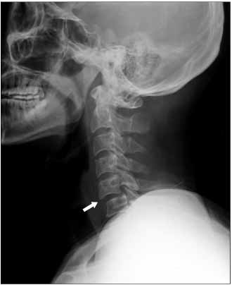
Fig. 1 Cervical spine lateral radiograph shows partial anterior translation of the body of the fifth cervical vertebra.
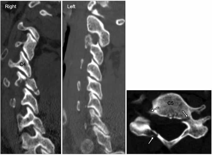
Fig. 2 CT scans show the bilateral fracture of the arch at the fifth cervical vertebra, the ipsilateral fractures of the pedicle and lamina on the right, and the fracture of the pedicle on the left.
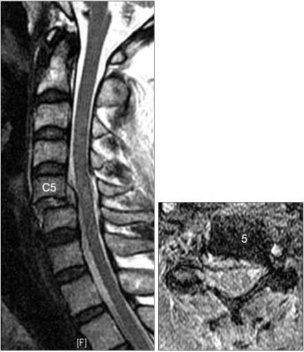
Fig. 3 MR images shows dislocation of the right facet joint at C4-5 and disc herniation at the C5-6 level with partial anterior displacement of the C5 body.
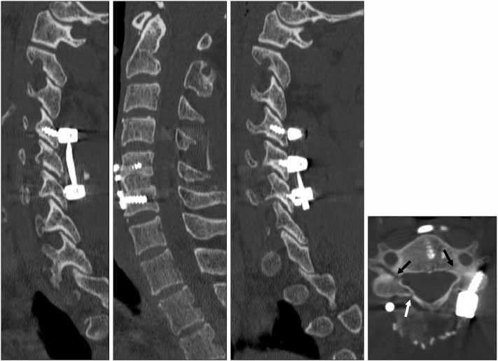
Fig. 4 Postoperative CT scans show good alignment of the cervical spine with a good reduction of the C4-5 facet and C5 body.

Fig. 5 One-year follow up radiograph of the cervical spine shows good fusion and good alignment.

Fig. 6 Preoperative cervical spine lateral radiography and MRI shows partial anterior translation of the 6th cervical vertebral body. There is no evidence of a disc or ligamentous injury.
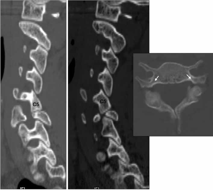
Fig. 7 CT scans show partial anterior translation of the sixth cervical vertebra body, bilateral dislocation of C5-6 facet joints, and subluxation of the C6-7 facet joints. Bilateral fracture of the arch at the sixth cervical vertebra at the isthmic portion between the lateral mass and pedicle is shown on the axial image.
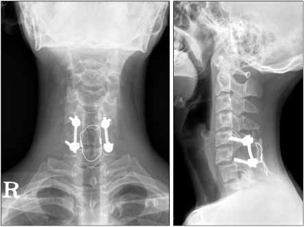
Fig. 8 One year follow up cervical spine radiographs show good union and good alignment.
Reference
-
1. Allen BL Jr, Ferguson RL, Lehman TR, O'Brien RP. A mechanistic classification of closed, indirect fractures and dislocations of the lower cervical spine. Spine. 1982. 7:1–27.
Article2. Bohlman HH. Acute fractures and dislocations of the cervical spine. An analysis of three hundred hospitalized patients and review of the literature. J Bone Joint Surg Am. 1979. 61:1119–1142.
Article3. Forsyth HF. Extension injuries of the cervical spine. J Bone Joint Surg Am. 1964. 46:1792–1797.
Article4. Fuentes JM, Benezech J, Lussiez B, Vlahovitch B. Fracture-separation of the articular process of the lower cervical spine. Its relation to fracture-dislocation in hyperextension. Rev Chir Orthop Reparatrice Appar Mot. 1986. 72:435–440.
- Full Text Links
-
- Actions
-
Cited
- CITED
-
- Close
- Share
-
- Similar articles
-
- Relationship between Soft Tissue Damages and Spinal Cord Injury in Lower Cervical Spine Trauma
- Delayed or Missed Diagnosis of Cervical Instability after Traumatic Injury: Usefulness of Dynamic Flexion and Extension Radiographs
- Squamous Cell Carcinoma of the Cervix with Intraepithelial Extension to the Endometrium: A Case Report
- Soft Tissue Damage in Cervical Spine Extension Injury
- A Clinical Analysis of Traumatic Cervical Spine Injuries
