J Korean Orthop Assoc.
2008 Oct;43(5):579-587. 10.4055/jkoa.2008.43.5.579.
Factors Affecting Segmental Motion of Lumbar Total Disc Replacement
- Affiliations
-
- 1Department of Orthopaedic Surgery,Samsung Medical Center, Sungkyunkwan University School of Medicine, Seoul, Korea. osfracture@naver.com
- 2Department of Radiology and Center for Imaging Science,Samsung Medical Center, Sungkyunkwan University School of Medicine, Seoul, Korea.
- KMID: 2186419
- DOI: http://doi.org/10.4055/jkoa.2008.43.5.579
Abstract
-
PURPOSE: To assess factors significantly affecting the range of motion of the lumbar spine at the operated segment following total disc replacement (TDR) arthroplasty.
MATERIALS AND METHODS
Thirty-six patients (15 men and 21 women) who received lumbar TDR at a single level using Prodisc II (Spine Solutions Inc, New York, NY USA) were included in this study. The study included 23 cases at L4-5 and 13 cases at L5-S1. The average patient age was 43.6 years (range, 23-59 years) and the minimum follow-up was 24 months (range, 24-61 months). Two independent observers measured radiological parameters preoperatively, at 3 months postoperatively, and at the final follow-up. These parameters included disc height, affected level segmental range of motion (ROM) and prosthesis position and height. A radiologist independently measured facet joint degeneration and the fat contents of the paraspinal muscles on preoperative MR images. Clinical results were evaluated using the Oswestry Disability Index (ODI) and the Visual Analogue Scale (VAS).
RESULTS
Segmental ROM was well preserved at the final follow-up (preoperative, 11.3 degrees; 3 months postoperative 13.2 degrees; final follow up, 13.1 degrees). The factors found to affect segmental ROM significantly at the final follow-up were the preoperative ROM, preoperative disc height, disc height increment ratio and a history of previous back surgery on the affected disc (p<0.05). The VAS significantly improved in patients with increased segmental ROM at the operated level (p<0.05).
CONCLUSION
Statistical analysis showed that the factors affecting segmental ROM were the preoperative ROM, preoperative disc height, disc height increment ratio, and a history of previous back surgery on the affected disc. However, further effort needs to be directed towards an evaluation of a larger number of patients with a longer follow-up.
MeSH Terms
Figure
-

Fig. 1 Flexion-extension radiographs of a patient who underwent TDR implantation at L4-L5. The TDR ROM was measured using the Cobb method with the keels as radiographic landmarks in flexion and extension X-rays.
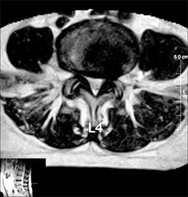
Fig. 2 Muscle fatty degeneration grade 2, right facet joint degeneration grade 1 and left grade 3.
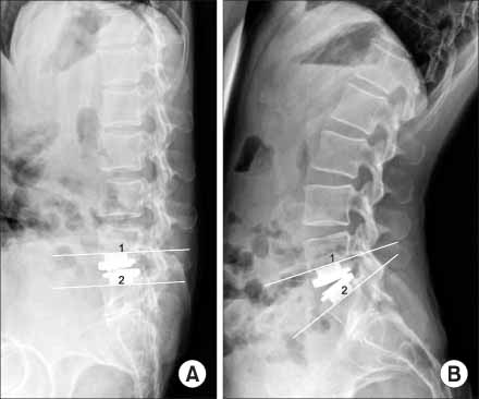
Fig. 3 Lateral flexion (A) and extension (B) radiographs at 4-years of a 40-year-old woman with the Prodisc II applied to L5-S1. Segmental ROM was 18°. Lumbarization of S1.
Reference
-
1. Auerbach JD, Wills BP, McIntosh TC, Balderston RA. Evaluation of spinal kinematics following lumbar total disc replacement and circumferential fusion using in vivo fluoroscopy. Spine. 2007. 32:527–536.
Article2. Bertagnoli R, Kumar S. Indications for full prosthetic disc arthroplasty: a correlation of clinical outcome against a variety of indications. Eur Spine J. 2002. 11:Suppl 2. S131–S136.
Article3. Blasier RD, Monson RC. Acquired spondylolysis after posterolateral spinal fusion. J Pediatr Orthop. 1987. 7:215–217.
Article4. Brunet JA, Wiley JJ. Acquired spondylolysis after spinal fusion. J Bone Joint Surg Br. 1984. 66:720–724.
Article5. Büttner-Janz K, Schellnack K, Zippel H. An alternative treatment strategy in lumbar intervertebral disk damage using an SB Charité modular type intervertebral disk endoprosthesis. Z Orthop Ihre Grenzgeb. 1987. 125:1–6.6. Chazal J, Tanguy A, Bourges M, et al. Biomechanical properties of spinal ligaments and a histological study of the supraspinal ligament in traction. J Biomech. 1985. 18:167–176.
Article7. Chung SS, Lee CS, Kang CS. Lumbar total disc replacement using ProDisc II: a prospective study with a 2-year minimum follow-up. J Spinal Disord Tech. 2006. 19:411–415.8. Cinotti G, David T, Postacchini F. Results of disc prosthesis after a minimum follow-up period of 2 years. Spine. 1996. 21:995–1000.
Article9. David T. Long-term results of one-level lumbar arthroplasty: minimum 10-year follow-up of the CHARITE artificial disc in 106 patients. Spine. 2007. 32:661–666.10. Eijkelkamp MF, van Donkelaar CC, Veldhuizen AG, van Horn JR, Huyghe JM, Verkerke GJ. Requirements for an artificial intervertebral disc. Int J Artif Organs. 2001. 24:311–321.
Article11. Errico TJ. Lumbar disc arthroplasty. Clin Orthop Relat Res. 2005. 435:106–117.
Article12. Goutallier D, Postel JM, Bernageau J, Lavau L, Voisin MC. Fatty muscle degeneration in cuff ruptures. Pre- and postoperative evaluation by CT scan. Clin Orthop Relat Res. 1994. 304:78–83.13. Harris RI, Wiley JJ. Acquired spondylolysis as a sequel to spine fusion. J Bone Joint Surg Am. 1963. 45:1159–1170.
Article14. Huang RC, Girardi FP, Cammisa FP Jr, Lim MR, Tropiano P, Marnay T. Correlation between range of motion and outcome after lumbar total disc replacement: 8.6-year follow-up. Spine. 2005. 30:1407–1411.
Article15. Huang RC, Girardi FP, Cammisa FP Jr, Tropiano P, Marnay T. Long-term flexion-extension range of motion of the prodisc total disc replacement. J Spinal Disord Tech. 2003. 16:435–440.
Article16. Huang RC, Tropiano P, Marnay T, Girardi FP, Lim MR, Cammisa FP Jr. Range of motion and adjacent level degeneration after lumbar total disc replacement. Spine J. 2006. 6:242–247.
Article17. Jackson RK, Boston DA, Edge AJ. Lateral mass fusion. A prospective study of a consecutive series with long-term follow-up. Spine. 1985. 10:828–832.18. Kettler A, Wilke HJ. Review of existing grading systems for cervical or lumbar disc and facet joint degeneration. Eur Spine J. 2006. 15:705–718.
Article19. Kiviluoto O, Santavirta S, Salenius P, Morri P, Pylkkänen P. Postero-lateral spine fusion. A 1-4-year follow-up of 80 consecutive patients. Acta Orthop Sca. d. 1985. 56:152–154.20. Le Huec JC, Basso Y, Aunoble S, Friesem T, Bruno MB. Influence of facet and posterior muscle degeneration on clinical results of lumbar total disc replacement: two-year follow-up. J Spinal Disord Tech. 2005. 18:219–223.21. Lee CK, Langrana NA, Parsons JR, Zimmerman MC. Development of a prosthetic intervertebral disc. Spine. 1991. 16:Suppl 6. S253–S255.
Article22. Lim MR, Girardi FP, Zhang K, Huang RC, Peterson MG, Cammisa FP Jr. Measurement of total disc replacement radiographic range of motion: a comparison of two techniques. J Spinal Disord Tech. 2005. 18:252–256.23. Link HD. History, design and biomechanics of the LINK SB charité artificial disc. Eur Spine J. 2002. 11:Suppl 2. S98–S105.
Article24. McAfee PC, Cunningham B, Holsapple G, et al. A prospective, randomized, multicenter Food and Drug Administration investigational device exemption study of lumbar total disc replacement with the CHARITE artificial disc versus lumbar fusion: part II: evaluation of radiographic outcomes and correlation of surgical technique accuracy with clinical outcomes. Spine. 2005. 30:1576–1583.25. Mengiardi B, Schmid MR, Boos N, et al. Fat content of lumbar paraspinal muscles in patients with chronic low back pain and in asymptomatic volunteers: quantification with MR spectroscopy. Radiology. 2006. 240:786–792.
Article26. O'Beirne J, O'Neill D, Gallagher J, Williams DH. Spinal fusion for back pain: a clinical and radiological review. J Spinal Disord. 1992. 5:32–38.27. Putzier M, Funk JF, Schneider SV, et al. Charité total disc replacement--clinical and radiographical results after an average follow-up of 17 years. Eur Spine J. 2006. 15:183–195.
Article28. Regan JJ. Clinical results of charité lumbar total disc replacement. Orthop Clin North Am. 2005. 36:323–340.
Article29. Tropiano P, Huang RC, Girardi FP, Marnay T. Lumbar disc replacement: preliminary results with ProDisc II after a minimum follow-up period of 1 year. J Spinal Disord Tech. 2003. 16:362–368.30. Weishaupt D, Zanetti M, Boos N, Hodler J. MR imaging and CT in osteoarthritis of the lumbar facet joints. Skeletal Radiol. 1999. 28:215–219.
Article31. West JL 3rd, Bradford DS, Ogilvie JW. Results of spinal arthrodesis with pedicle screw-plate fixation. J Bone Joint Surg Am. 1991. 73:1179–1184.
Article32. Wong KW, Leong JC, Chan MK, Luk KD, Lu WW. The flexion-extension profile of lumbar spine in 100 healthy volunteers. Spine. 2004. 29:1636–1641.
Article
- Full Text Links
-
- Actions
-
Cited
- CITED
-
- Close
- Share
-
- Similar articles
-
- Lumbar Total Disc Replacement
- Current Status of Lumbar Total Disc Replacement (TDR)
- Motion Study in the Fused Lumbar Spine
- Lumbar Disc Degeneration and Segmental Instability: A Comparison of Magnetic Resonance Images and Plain Radiographs
- Total Cervical Disc Replacement using Artificial Disc in Cervical Disc Herniations
