Factors for Recurrence in Pigmented Villonodular Synovitis of Large Joints
- Affiliations
-
- 1Department of Orthopedic Surgery, Seoul National University College of Medicine, Seoul, Korea. hankim@snu.ac.kr
- KMID: 2186414
- DOI: http://doi.org/10.4055/jkoa.2008.43.5.544
Abstract
-
PURPOSE: Pigmented villonodular synovitis (PVNS) is a rare proliferative disorder of the synovium, and this can affect the joints, tendon sheaths or bursae. PVNS is histologically benign, but it has a high propensity for local recurrence. The purpose of this study is to identify the clinical and pathological factors that are associated with local recurrence of PVNS.
MATERIALS AND METHODS
Fifty-one patients with biopsy-proven PVNS were retrospectively reviewed. There were 20 men and 31 women with an average age of 34 years (range: 12-73). The average follow-up period was 4.1 years (range: 1-25 years). All lesions were located in large joints (knee 25, ankle 11, hip 7, foot 5, wrist 2, elbow 1). Of the 51 lesions in the large joints, 39 were the diffuse type and 12 were the localized type. The initial clinical presentation was pain or a painful mass in 32 patients and a painless mass in 19 patients. Complete surgical removal of the lesion was performed in 39 cases, whereas incomplete excision was performed in 12. No adjuvant therapy was given in any cases.
RESULTS
Sixteen local recurrences (31%) developed at an average of 24 months (range, 4-96). Factors related to local recurrence in the large joints were incomplete surgical removal (p < 0.001), diffuse type of the lesion (p=0.049) and the presence of bone erosion (p=0.037)
CONCLUSION
In cases of PVNS in large joints, the factors that increased the local recurrence rate were incomplete surgical removal, diffuse type lesion and the presence of bone erosion.
MeSH Terms
Figure
-

Fig. 1 A representative histological specimen is shown. Note the accumulation of mononuclear cells with interspersed giant cells and lipid laden macrophages (H&E stain, ×400).

Fig. 2 Representative MRI images of the two types of PVNS are presented. (A) A localized type of PVNS is located in the posterior aspect of the distal femur. (B) A diffuse type of PVNS is seen at the posterior part of the ankle.

Fig. 3 A 14-year-old girl was diagnosed with PVNS in her left hip joint. (A) Plain radiography suggests bone erosion at the femoral head and acetabulum of the left hip joint (arrow). (B) MRI image shows the tumor eroding into the acetabulum and the femoral head.
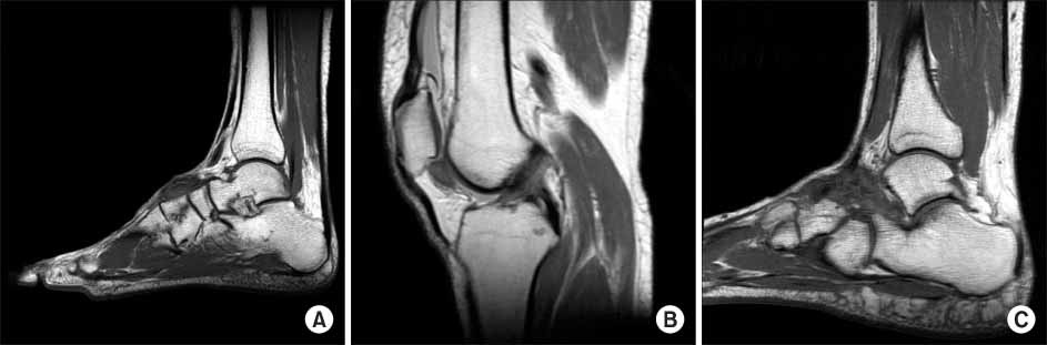
Fig. 4 Representative MRI images of PVNS according the location of the lesion are presented. (A) Extraarticular location, (B) intraarticular location, (C) mixed location.

Fig. 5 Kaplan-Meier curve showing the risk of recurrence is illustrated. The recurrence-free survival rate was 90% and 60% at postoperative 1 and 5 years respectively.
Reference
-
1. Byers PD, Cotton RE, Deacon OW, et al. The diagnosis and treatment of pigmented villonodular synovitis . J Bone Joint Surg Br. 1968. 50:290–305.
Article2. Chassaignac M. Cancer de la gaine des tendons. Gaz Hop Civ Milit. 1852. 57:185–186.3. Chiari C, Pirich C, Brannath W, Kotz R, Trieb K. What affects the recurrence and clinical outcome of pigmented villonodular synovitis? Clin Orthop Relat Res. 2006. 450:172–178.
Article4. Chin KR, Barr SJ, Winalski C, Zurakowski D, Brick GW. Treatment of advanced primary and recurrent diffuse pigmented villonodular synovitis of the knee. J Bone Joint Surg Am. 2002. 84:2192–2202.
Article5. Choong PF, Willén H, Nilbert M, et al. Pigmented villonodular synovitis. Monoclonality and metastasis--a case for neoplastic origin? Acta Orthop Scand. 1995. 66:64–68.
Article6. Chung SM, Janes JM. Diffuse pigmented villonodular synovitis of the hip joint. Review of the literature and report of four cases. J Bone Joint Surg Am. 1965. 47:293–303.7. de Visser E, Veth RP, Pruszczynski M, Wobbes T, Van de Putte LB. Diffuse and localized pigmented villonodular synovitis: evaluation of treatment of 38 patients. Arch Orthop Trauma Surg. 1999. 119:401–404.
Article8. Dürr HR, Stäbler A, Maier M, Refior HJ. Pigmented villonodular synovitis. Review of 20 cases. J Rheumatol. 2001. 28:1620–1630.9. Flandry FC, Hughston JC, Jacobson KE, Barrack RL, McCann SB, Kurtz DM. Surgical treatment of diffuse pigmented villonodular synovitis of the knee. Clin Orthop Relat Res. 1994. 183–192.
Article10. Fletcher JA, Henkle C, Atkins L, Rosenberg AE, Morton CC. Trisomy 5 and trisomy 7 are nonrandom aberrations in pigmented villonodular synovitis: confirmation of trisomy 7 in uncultured cells. Genes Chromosomes Cancer. 1992. 4:264–266.
Article11. Granowitz SP, D'Antonio J, Mankin HL. The pathogenesis and long-term end results of pigmented villonodular synovitis. Clin Orthop Relat Res. 1976. 335–351.12. Hwang DS, Lee CH, Yang JH, Kang TH. Pigmented villonodular synovitis of the ankle joint. J Korean Orthop Assoc. 2006. 41:905–910.
Article13. Jaffe H, Lichtenstein L, Sutro C. Pigmented villonodular synovitis, bursitis and tenosynovitis. A discussion of synovial and bursal equivalents of tenosynovial lesion commonly denoted as xanthoma, xanthogranuloma, giant cell tumor, or myeloplaxoma of tendon sheath, with some considerations of this tendon sheath lesion itself. Arch Pathol. 1941. 31:731–765.14. Johansson JE, Ajjoub S, Coughlin LP, Wener JA, Cruess RL. Pigmented villonodular synovitis of joints. Clin Orthop Relat Res. 1982. 159–166.
Article15. Kim KT, Kim CH, Lee MJ. Arthroscopic treatment for the pigmented villonodular synovitis in the knee. The Journal of Korean Arthroscopy Society. 2001. 5:111–115.16. Kim RS, Kim CS, Park HW, Park JH. Clinical features of diffuse pigmented villonodular synovitis in the knee joint. J Korean Orthop Assoc. 2002. 37:215–219.
Article17. Kotwal PP, Gupta V, Malhotra R. Giant-cell tumour of the tendon sheath. Is radiotherapy indicated to prevent recurrence after surgery? J Bone Joint Surg Br. 2000. 82:571–573.18. Kramer D, Frassica F, Cosgarea A. Total arthroscopic synovectomy for pigmented villonodular synovitis of the knee. Techniques Knee Surg. 2004. 3:36–45.
Article19. Mendenhall WM, Mendenhall CM, Reith JD, Scarborough MT, Gibbs CP, Mendenhall NP. Pigmented villonodular synovitis. Am J Clin Oncol. 2006. 29:548–550.
Article20. Mohr W. Pigmented villonodular synovitis--a review with reference to 166 cases . Pathologe. 1992. 13:314–321.21. O'Connell JX. Pathology of the synovium. Am J Clin Pathol. 2000. 114:773–784.22. Ofluoglu O. Pigmented villonodular synovitis. Orthop Clin North Am. 2006. 37:23–33.
Article23. Ogilvie-Harris DJ, McLean J, Zarnett ME. Pigmented villonodular synovitis of the knee. The results of total arthroscopic synovectomy, partial, arthroscopic synovectomy, and arthroscopic local excision. J Bone Joint Surg Am. 1992. 74:119–123.
Article24. Rao AS, Vigorita VJ. Pigmented villonodular synovitis (giant-cell tumor of the tendon sheath and synovial membrane). A review of eighty-one cases. J Bone Joint Surg Am. 1984. 66:76–94.
Article25. Ray RA, Morton CC, Lipinski KK, Corson JM, Fletcher JA. Cytogenetic evidence of clonality in a case of pigmented villonodular synovitis. Cancer. 1991. 67:121–125.
Article26. Schwartz HS, Unni KK, Pritchard DJ. Pigmented villonodular synovitis. A retrospective review of affected large joints. Clin Orthop Relat Res. 1989. 243–255.
- Full Text Links
-
- Actions
-
Cited
- CITED
-
- Close
- Share
-
- Similar articles
-
- Localized Pigmented Villonodular Synovitis of the Posterior Compartment of the Knee: A Case Report
- Arthroscopic Treatment of Pigmented Villonodular Synovitis of the Shoulder: A Case Report
- Localized Pigmented Villonodular Synovitis with Recurrent Subluxation of the Patella: A Case Report
- Pigmented Villonodular Synovitis of the Spine: A Case Report
- Total Ankle Replacement in Pigmented Villonodular Synovitis of Ankle Joint (A Case Report)
