Short to Midterm Results of Bernese Periacetabular Osteotomy for Hip Dysplasia
- Affiliations
-
- 1Department of Orthopedic Sugery, Seoul National University College of Medicine, Seoul, Korea. oskim@snu.ac.kr
- KMID: 2185277
- DOI: http://doi.org/10.4055/jkoa.2013.48.6.433
Abstract
- PURPOSE
The goal of this study was to analyze the short to midterm results of Bernese periacetabular osteotomy for the patient with hip dysplasia.
MATERIALS AND METHODS
Fourteen Bernese periacetabular osteotomies were performed in 13 patients and were followed-up for more than 2.5 years. All patients had been treated at Department of Orthopedic Surgery, Seoul National University Hospital from June 1998 to December 2007. We conducted an interview and asked the patients to complete the Harris hip score and pain visual analogue scale (VAS) score for clinical evaluation. Radiographic measurements included Tonnis osteoarthritis grade, center-edge angle of Wiberg, acetabular angle of Sharp, acetabular depth index, acetabular head index.
RESULTS
Mean Harris hip score improved from 63.8 points preoperatively to 82.9 points postoperatively and pain VAS score improved from 7.6 points preoperatively to 0.8 points postoperatively. Based on Tonnis osteoarthritis grade, progression of osteoarthritis was found in 4 hips. Radiologically, center-edge angle of Wiberg, acetabular angle of Sharp, acetabular depth index and acetabular head index improved. Complications included superficial skin infection in one hip and non-union of pubic ramus in one. One patient had metal hypersensitivity to cortical screws and fracture of posterior column.
CONCLUSION
The short to midterm results show Bernese periacetabular osteotomy is a satisfactory technique for treating early and mild hip-osteoarthritis patients with hip dysplasia clinically and radiographically.
Figure
-
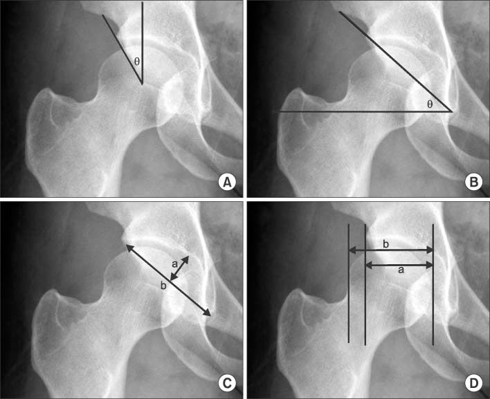
Figure 1 The diagram shows radiographic measurements. (A) Center-edge angle of Wiberg, (B) acetabular angle of Sharp, (C) acetabular depth index: a/b×1,000, (D) acetabular head index: a/b×100.

Figure 2 The flow chart shows the postoperative osteoarthritic changes depending on the preoperative Tönnis osteoarthritis grade.

Figure 3 Radiographs are obtained from a 50-year-old female patient who underwent a Bernese periacetabular osteotomy. (A) Preoperative radiograph (anteroposterior). (B) Postoperative radiogarphs taken at 3 years after surgery (anteroposterior & oblique). (C) Radiogarph at 8 years after surgery (anteroposterior & oblique). (D) Radiograph at 13 years after surgery (anteroposterior).

Figure 4 Anteroposterior radiographs are showing the hips and pelvis of a 47-year-old woman who was managed with a Bernese periacetabular osteotomy. (A) Preoperative radiograph. (B) Postoperative radiogarphs taken at 1 year after surgery of the right hip. (C) Immediate postoperative radiogarphs of the left hip. (D) Radiograph at 3 years after surgery of the left hip.
Reference
-
1. Leunig M, Siebenrock KA, Ganz R. Rationale of periacetabular osteotomy and background work. Instr Course Lect. 2001; 50:229–238.
Article2. Murphy SB, Kijewski PK, Millis MB, Harless A. Acetabular dysplasia in the adolescent and young adult. Clin Orthop Relat Res. 1990; 261:214–223.
Article3. Murphy SB, Ganz R, Müller ME. The prognosis in untreated dysplasia of the hip. A study of radiographic factors that predict the outcome. J Bone Joint Surg Am. 1995; 77:985–989.
Article4. Staheli LT, Chew DE. Slotted acetabular augmentation in childhood and adolescence. J Pediatr Orthop. 1992; 12:569–580.
Article5. Cooperman DR, Wallensten R, Stulberg SD. Acetabular dysplasia in the adult. Clin Orthop Relat Res. 1983; 175:79–85.
Article6. Ganz R, Klaue K, Vinh TS, Mast JW. A new periacetabular osteotomy for the treatment of hip dysplasias. Technique and preliminary results. Clin Orthop Relat Res. 1988; 232:26–36.
Article7. Sharp IK. Acetabular dysplasia. the acetabular angle. J Bone Joint Surg Br. 1961; 43:268–272.8. Hussell JG, Mast JW, Mayo KA, Howie DW, Ganz R. A comparison of different surgical approaches for the periacetabular osteotomy. Clin Orthop Relat Res. 1999; 363:64–72.
Article9. Millis MB, Poss R, Murphy SB. Osteotomies of the hip in the prevention and treatment of osteoarthritis. Instr Course Lect. 1992; 41:145–154.10. Ninomiya S, Tagawa H. Rotational acetabular osteotomy for the dysplastic hip. J Bone Joint Surg Am. 1984; 66:430–436.
Article11. Peters CL, Erickson JA, Hines JL. Early results of the Bernese periacetabular osteotomy: the learning curve at an academic medical center. J Bone Joint Surg Am. 2006; 88:1920–1926.
Article12. Hussell JG, Rodriguez JA, Ganz R. Technical complications of the Bernese periacetabular osteotomy. Clin Orthop Relat Res. 1999; 363:81–92.
Article13. Chang JS, Kwon KD, Shon HC. Bernese periacetabular osteotomy using dual approaches for hip dysplasias. J Korean Orthop Assoc. 2002; 37:226–232.
Article14. Park YS, Moon YW, Lim SJ, Park JC, Son MS. Short-term follow-up results of periacetabular osteotomy for hip dysplasia. J Korean Hip Soc. 2009; 21:156–161.
Article15. Siebenrock KA, Leunig M, Ganz R. Periacetabular osteotomy: the Bernese experience. Instr Course Lect. 2001; 50:239–245.
Article16. Matheney T, Kim YJ, Zurakowski D, Matero C, Millis M. Intermediate to long-term results following the Bernese periacetabular osteotomy and predictors of clinical outcome. J Bone Joint Surg Am. 2009; 91:2113–2123.
Article17. Naito M, Shiramizu K, Akiyoshi Y, Ezoe M, Nakamura Y. Curved periacetabular osteotomy for treatment of dysplastic hip. Clin Orthop Relat Res. 2005; 433:129–135.
Article18. Steppacher SD, Tannast M, Ganz R, Siebenrock KA. Mean 20-year followup of Bernese periacetabular osteotomy. Clin Orthop Relat Res. 2008; 466:1633–1644.
Article
- Full Text Links
-
- Actions
-
Cited
- CITED
-
- Close
- Share
-
- Similar articles
-
- Bernese Periacetabular Osteotomy for Treatment of Acetabular Dysplasia
- Bernese Periacetabular Osteotomy Using Dual Approaches for Hip Dysplasias
- Pelvic Osteotomy in Adults
- Osteotomies Around the Hip Joint
- Periacetabular Osteotomy for Treating Dysplastic Hips That Were Misdiagnosed as Having Acetabular Bone Tumor: Report of Two Cases
