Granular Cell Tumor in the Sartorius Muscle
- Affiliations
-
- 1Department of Orthopaedic Surgery, Soonchunhyang University Hospital Cheonan, Cheonan, Korea. huuy@schmc.ac.kr
- KMID: 2185258
- DOI: http://doi.org/10.4055/jkoa.2014.49.1.69
Abstract
- Granular cell tumor, a soft tissue neoplasm that originates in the nervous system, is a very unusual tumor. Granular cell tumor appears as a solitary painless lesion, which can arise at virtually any body site, but is mainly found on the skin, oral cavity, respiratory tract or digestive tract. However, an intramuscular granular cell tumor is very rare. We report on a case of a granular cell tumor in the sartorius muscle in a 71-year-old male patient along with a review of the literature.
Keyword
MeSH Terms
Figure
-
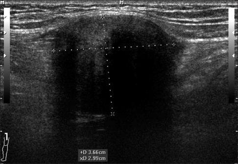
Figure 1 Preoperative ultrasound image shows a low echoic mass measuring 6×4 cm in size with a central high echoic lesion in the sartorius muscle.
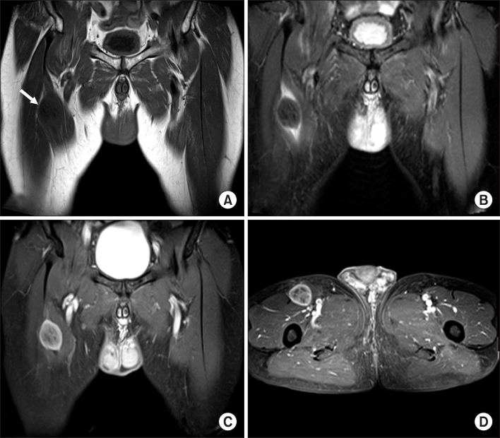
Figure 2 (A) Coronal non-contrast T1-weighted image of the pelvis shows a low signal intensity mass within the mid portion of the sartorius muscle measuring 6×5×4 cm in size (arrow). (B) Coronal non-contrast T2-weighted image shows a low signal intensity mass with peripheral ovoid high signal intensity. (C) T1-weighted enhanced image shows a heterogeneous enhancing mass with a clear margin. (D) Axial T1-weighted enhanced image shows a mass in the mid portion of the sartorius muscle.

Figure 3 (A) The submitted specimen was comprised of a well demarcated ovoid mass measuring 4×5×6 cm in size. (B) On sectioning, a pale yellow surface with firm consistency was observed.
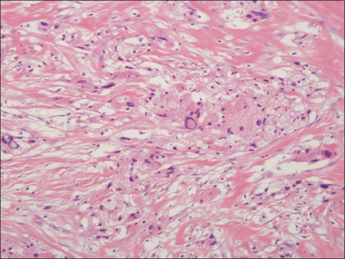
Figure 4 Histologic examination showed the characteristic appearance of nests of large polyhedral cells with abundant granular cytoplasm (H&E, ×400).
Reference
-
1. Khansur T, Balducci L, Tavassoli M. Granular cell tumor. Clinical spectrum of the benign and malignant entity. Cancer. 1987; 60:220–222.
Article2. Arai E, Nishida Y, Tsukushi S, Sugiura H, Ishiguro N. Intramuscular granular cell tumor in the lower extremities. Clin Orthop Relat Res. 2010; 468:1384–1389.
Article3. Fanburg-Smith JC, Meis-Kindblom JM, Fante R, Kindblom LG. Malignant granular cell tumor of soft tissue: diagnostic criteria and clinicopathologic correlation. Am J Surg Pathol. 1998; 22:779–794.4. Elkousy H, Harrelson J, Dodd L, Martinez S, Scully S. Granular cell tumors of the extremities. Clin Orthop Relat Res. 2000; 380:191–198.
Article5. Thacker MM, Humble SD, Mounasamy V, Temple HT, Scully SP. Case report. Granular cell tumors of extremities: comparison of benign and malignant variants. Clin Orthop Relat Res. 2007; 455:267–273.6. Ordóñez NG. Granular cell tumor: a review and update. Adv Anat Pathol. 1999; 6:186–203.
Article7. Lack EE, Worsham GF, Callihan MD, et al. Granular cell tumor: a clinicopathologic study of 110 patients. J Surg Oncol. 1980; 13:301–316.
Article8. Tsuchida T, Okada K, Itoi E, Sato T, Sato K. Intramuscular malignant granular cell tumor. Skeletal Radiol. 1997; 26:116–121.
Article9. Rosenthal SA, Livolsi VA, Turrisi AT 3rd. Adjuvant radiotherapy for recurrent granular cell tumor. Cancer. 1990; 65:897–900.
Article10. Blacksin MF, White LM, Hameed M, Kandel R, Patterson FR, Benevenia J. Granular cell tumor of the extremity: magnetic resonance imaging characteristics with pathologic correlation. Skeletal Radiol. 2005; 34:625–631.
Article
