Lateral Closing Wedge Osteotomy of Tibia for Degenerative Arthritis
- Affiliations
-
- 1Department of Orthopaedic Surgery, Seoul National University College of Medicine, Seoul, Korea. leemc@snu.ac.kr
- KMID: 2185214
- DOI: http://doi.org/10.4055/jkoa.2014.49.2.95
Abstract
- Proximal tibial osteotomy is an effective, well-established treatment for unicompartmental arthritic knee with varus or valgus deformity. Four basic types are commonly described: lateral closing wedge osteotomy, medial open wedge osteotomy, dome osteotomy, and medial opening hemicallotasis. The objective of this procedure is to realign the weight-bearing axis through the knee by redistributing the forces of weight to the less involved compartment of the knee. With thorough preoperative planning and careful selection of patients, optimal outcome can be expected with preservation of the patient's joint. In this article, we reviewed selection of patients, surgical planning, surgical technique, complications, pre- and post-operative change in mechanics, and long term surgical outcome of closing wedge osteotomy. Optimal outcome is expected in patients with young age (younger than 60), stable knee, medially confined osteoarthritis, and good range of motion. According to the literature, average 10-year survival rate is expected to be 60% to 90%. Closing wedge osteotomy allows for rapid bone healing, early weight bearing, rehabilitation, and low rates of correction loss. Surgeons should keep in mind that optimal indication, preoperative planning, and use of safe operative technique are essential to achievement of best results.
MeSH Terms
Figure
-

Figure 1 Relationship between mechanical axis and anatomical axis in normal lower extremity. The mechanical axis is a line connecting the centers of the femur head and the tibiotalar joint, which passes through the center of the knee joint. It is in 3 degrees of valgus from the vertical axis of the body. Anatomical axis (femoral shaft axis) is in 6 degrees of valgus from the mechanical axis of the lower limb and 9 degrees of valgus from the true vertical axis of the body.
Figure 2 Progression of osteoarthritis in the varus knee (left to right). Varus deformity increases adduction moment of the knee during gait, which further increases medial tibiofemoral joint loading. Increased loading aggravates osteoarthritis and deformity. If lateral laxity accompanies, varus deformity can show rapid progress.

Figure 3 Preoperative simulation with a full-length standing radiograph. We simulate postoperative limb alignment according to the intersecting location of the mechanical axis. (A) This photograph shows preoperative alignment on the left extremity. (B) This photograph shows the mechanical axis that passes through the lateral tibial spine. (C) This photograph shows the Fujisawa point. We also measured anatomical axis and medial proximal tibial angle (MPTA) in each case. As the causes of varus deformity can differ, all of these variables should be considered together for development of an optimal results.
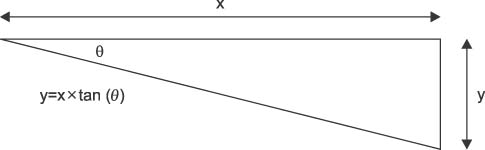
Figure 4 Calculation of wedge height usinga trigonometric method. Actual wedge height (y) is calculated with the desired angle of correction (θ) and actual tibial width (x). Actual tibial width should be measured at 2.0-2.5 cm distal to the joint line and should be normalized according to the amount of magnification.
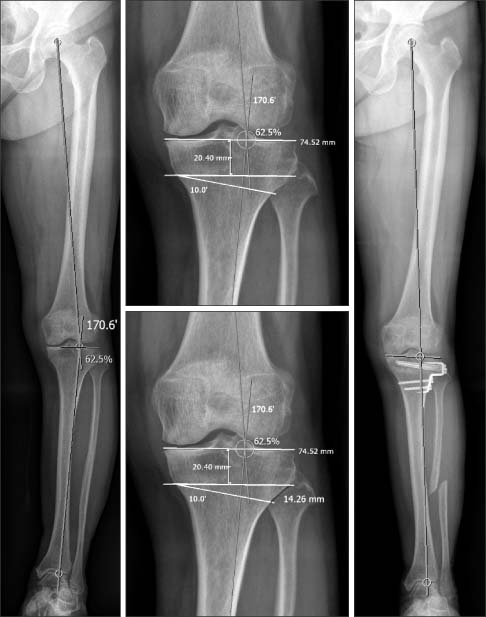
Figure 5 Calculation of wedge height using a picture archiving and communication system (PACS). Desired angle of correction is measured using teleradiography. In this figure, desired angle of correction is 10 degrees when the mechanical axis passes through the Fujisawa point. This angle is marked at 2 cm distal to the joint line and the wedge height can then be measured directly.
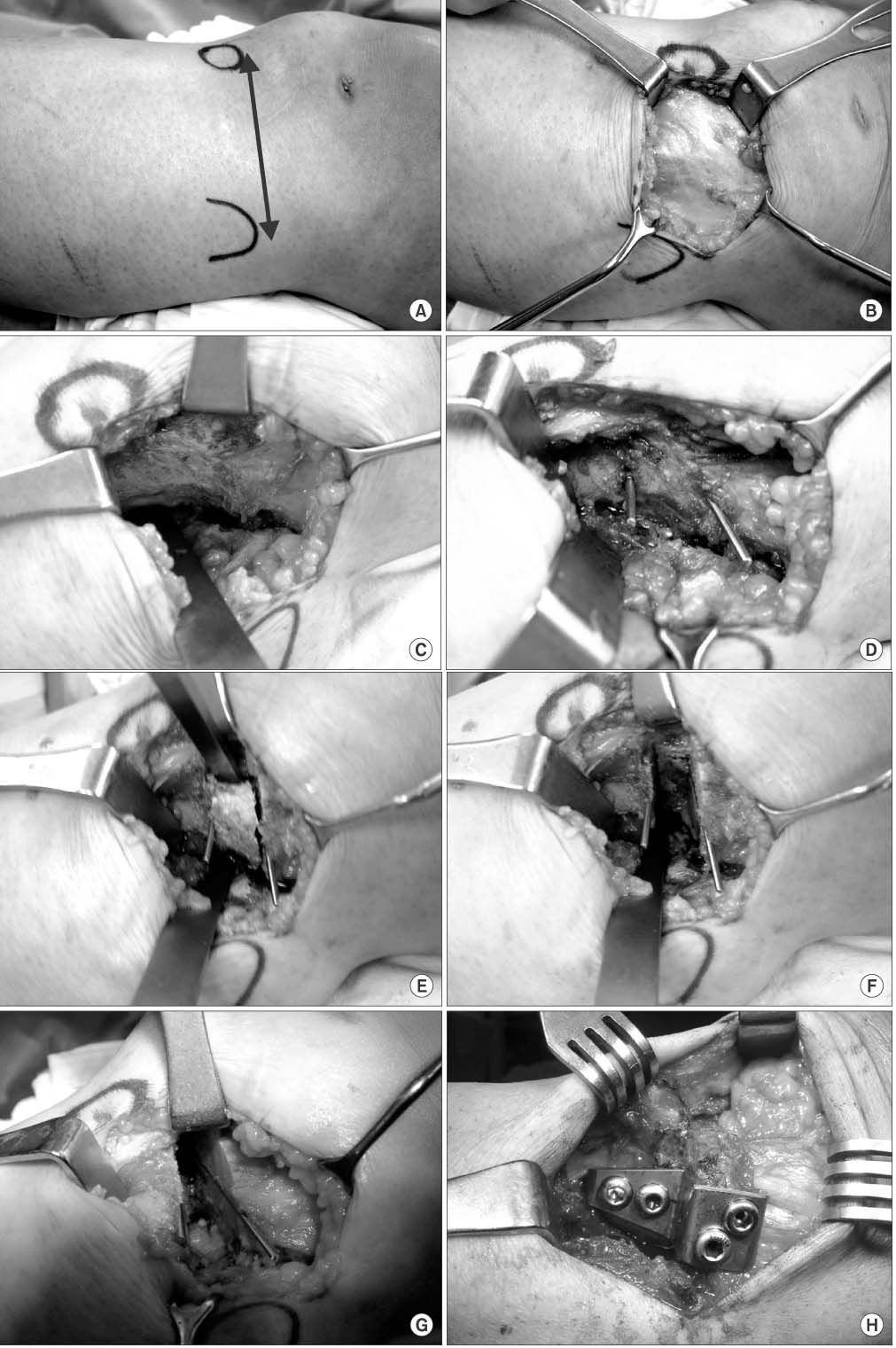
Figure 6 Intraoperative picture (see text for detailed description). (A) Transverse skin incision from tibial tuberosity to the fibular head. (B) Exposed fascia. (C) Subperiosteal stripping using an elevator. (D) Insertion of the guide pin under the c-arm. (E) Proximal wedge removal. (F) Distal wedge removal. (G) Application of gentle valgus force to make plastic deformation of the contralateral cortex. (H) Internal fixation.

Figure 7 Fixation device for closing wedge osteotomy. (A) Stepped staple (Coventry staple). (B) Blade plate. (C) L-plate (condylar buttress plate). (D) Screw fixation. (E) Rigid stepped plate.
Reference
-
1. Yasuda K, Majima T, Tsuchida T, Kaneda K. A ten- to 15-year follow-up observation of high tibial osteotomy in medial compartment osteoarthrosis. Clin Orthop Relat Res. 1992; 282:186–195.2. Bauer GC, Insall J, Koshino T. Tibial osteotomy in gonarthrosis (osteo-arthritis of the knee). J Bone Joint Surg Am. 1969; 51:1545–1563.3. Insall JN, Joseph DM, Msika C. High tibial osteotomy for varus gonarthrosis. A long-term follow-up study. J Bone Joint Surg Am. 1984; 66:1040–1048.4. Naudie D, Bourne RB, Rorabeck CH, Bourne TJ. The Install Award. Survivorship of the high tibial valgus osteotomy. A 10- to -22-year followup study. Clin Orthop Relat Res. 1999; 367:18–27.5. Coventry MB. Osteotomy of the upper portion of the tibia for degenerative arthritis of the knee. A preliminary report. J Bone Joint Surg Am. 1965; 47:984–990.6. Rossi R, Bonasia DE, Amendola A. The role of high tibial osteotomy in the varus knee. J Am Acad Orthop Surg. 2011; 19:590–599.7. Lee DC, Byun SJ. High tibial osteotomy. Knee Surg Relat Res. 2012; 24:61–69.8. Aglietti P, Rinonapoli E, Stringa G, Taviani A. Tibial osteotomy for the varus osteoarthritic knee. Clin Orthop Relat Res. 1983; 176:239–251.9. Rudan JF, Simurda MA. High tibial osteotomy. A prospective clinical and roentgenographic review. Clin Orthop Relat Res. 1990; 255:251–256.10. Dugdale TW, Noyes FR, Styer D. Preoperative planning for high tibial osteotomy. The effect of lateral tibiofemoral separation and tibiofemoral length. Clin Orthop Relat Res. 1992; 274:248–264.11. Marti CB, Gautier E, Wachtl SW, Jakob RP. Accuracy of frontal and sagittal plane correction in open-wedge high tibial osteotomy. Arthroscopy. 2004; 20:366–372.12. Ryan PJ. Bone SPECT of the knees. Nucl Med Commun. 2000; 21:877–885.13. Kettelkamp DB, Wenger DR, Chao EY, Thompson C. Results of proximal tibial osteotomy. The effects of tibiofemoral angle, stance-phase flexion-extension, and medial-plateau force. J Bone Joint Surg Am. 1976; 58:952–960.14. Hsu RW, Himeno S, Coventry MB, Chao EY. Normal axial alignment of the lower extremity and load-bearing distribution at the knee. Clin Orthop Relat Res. 1990; 255:215–227.15. Coventry MB, Ilstrup DM, Wallrichs SL. Proximal tibial osteotomy. A critical long-term study of eighty-seven cases. J Bone Joint Surg Am. 1993; 75:196–201.16. Myrnerts R. Optimal correction in high tibial osteotomy for varus deformity. Acta Orthop Scand. 1980; 51:689–694.17. Hernigou P, Medevielle D, Debeyre J, Goutallier D. Proximal tibial osteotomy for osteoarthritis with varus deformity. A ten to thirteen-year follow-up study. J Bone Joint Surg Am. 1987; 69:332–354.18. Fujisawa Y, Masuhara K, Shiomi S. The effect of high tibial osteotomy on osteoarthritis of the knee. An arthroscopic study of 54 knee joints. Orthop Clin North Am. 1979; 10:585–608.19. Ogata K, Yoshii I, Kawamura H, Miura H, Arizono T, Sugioka Y. Standing radiographs cannot determine the correction in high tibial osteotomy. J Bone Joint Surg Br. 1991; 73:927–931.20. Prodromos CC, Andriacchi TP, Galante JO. A relationship between gait and clinical changes following high tibial osteotomy. J Bone Joint Surg Am. 1985; 67:1188–1194.21. Naudie DD, Amendola A, Fowler PJ. Opening wedge high tibial osteotomy for symptomatic hyperextension-varus thrust. Am J Sports Med. 2004; 32:60–70.22. Windsor RE, Insall JN, Vince KG. Technical considerations of total knee arthroplasty after proximal tibial osteotomy. J Bone Joint Surg Am. 1988; 70:547–555.23. Giffin JR, Vogrin TM, Zantop T, Woo SL, Harner CD. Effects of increasing tibial slope on the biomechanics of the knee. Am J Sports Med. 2004; 32:376–382.24. Lerat JL, Moyen B, Garin C, Mandrino A, Besse JL, Brunet-Guedj E. Anterior laxity and internal arthritis of the knee. Results of the reconstruction of the anterior cruciate ligament associated with tibial osteotomy. Rev Chir Orthop Reparatrice Appar Mot. 1993; 79:365–374.25. El-Azab H, Glabgly P, Paul J, Imhoff AB, Hinterwimmer S. Patellar height and posterior tibial slope after open- and closed-wedge high tibial osteotomy: a radiological study on 100 patients. Am J Sports Med. 2010; 38:323–329.26. Wright JM, Heavrin B, Begg M, Sakyrd G, Sterett W. Observations on patellar height following opening wedge proximal tibial osteotomy. Am J Knee Surg. 2001; 14:163–173.27. Westrich GH, Peters LE, Haas SB, Buly RL, Windsor RE. Patella height after high tibial osteotomy with internal fixation and early motion. Clin Orthop Relat Res. 1998; 354:169–174.28. Billings A, Scott DF, Camargo MP, Hofmann AA. High tibial osteotomy with a calibrated osteotomy guide, rigid internal fixation, and early motion. Long-term follow-up. J Bone Joint Surg Am. 2000; 82:70–79.29. Bae DK, Song SJ, Yoon KH. Total knee arthroplasty following closed wedge high tibial osteotomy. Int Orthop. 2010; 34:283–287.30. Gibson MJ, Barnes MR, Allen MJ, Chan RN. Weakness of foot dorsiflexion and changes in compartment pressures after tibial osteotomy. J Bone Joint Surg Br. 1986; 68:471–475.31. Marti CB, Jakob RP. Accumulation of irrigation fluid in the calf as a complication during high tibial osteotomy combined with simultaneous arthroscopic anterior cruciate ligament reconstruction. Arthroscopy. 1999; 15:864–866.32. Bettin D, Karbowski A, Schwering L, Matthiass HH. Time-dependent clinical and roentgenographical results of Coventry high tibial valgisation osteotomy. Arch Orthop Trauma Surg. 1998; 117:53–57.33. Aglietti P, Buzzi R, Vena LM, Baldini A, Mondaini A. High tibial valgus osteotomy for medial gonarthrosis: a 10- to 21-year study. J Knee Surg. 2003; 16:21–26.34. Matthews LS, Goldstein SA, Malvitz TA, Katz BP, Kaufer H. Proximal tibial osteotomy. Factors that influence the duration of satisfactory function. Clin Orthop Relat Res. 1988; 229:193–200.35. Sprenger TR, Doerzbacher JF. Tibial osteotomy for the treatment of varus gonarthrosis. Survival and failure analysis to twenty-two years. J Bone Joint Surg Am. 2003; 85:469–474.36. Parker DA, Viskontas DG. Osteotomy for the early varus arthritic knee. Sports Med Arthrosc. 2007; 15:3–14.37. Kessler OC, Jacob HA, Romero J. Avoidance of medial cortical fracture in high tibial osteotomy: improved technique. Clin Orthop Relat Res. 2002; 395:180–185.38. Tunggal JA, Higgins GA, Waddell JP. Complications of closing wedge high tibial osteotomy. Int Orthop. 2010; 34:255–261.39. Vainionpää S, Läike E, Kirves P, Tiusanen P. Tibial osteotomy for osteoarthritis of the knee. A five to ten-year follow-up study. J Bone Joint Surg Am. 1981; 63:938–946.40. Wootton JR, Ashworth MJ, MacLaren CA. Neurological complications of high tibial osteotomy--the fibular osteotomy as a causative factor: a clinical and anatomical study. Ann R Coll Surg Engl. 1995; 77:31–34.41. Ivarsson I, Myrnerts R, Gillquist J. High tibial osteotomy for medial osteoarthritis of the knee. A 5 to 7 and 11 year follow-up. J Bone Joint Surg Br. 1990; 72:238–244.42. Bauer T, Hardy P, Lemoine J, Finlayson DF, Tranier S, Lortat-Jacob A. Drop foot after high tibial osteotomy: a prospective study of aetiological factors. Knee Surg Sports Traumatol Arthrosc. 2005; 13:23–33.43. Kirgis A, Albrecht S. Palsy of the deep peroneal nerve after proximal tibial osteotomy. An anatomical study. J Bone Joint Surg Am. 1992; 74:1180–1185.44. van Raaij TM, Brouwer RW, de Vlieger R, Reijman M, Verhaar JA. Opposite cortical fracture in high tibial osteotomy: lateral closing compared to the medial opening-wedge technique. Acta Orthop. 2008; 79:508–514.45. Pape D, Adam F, Rupp S, Seil R, Kohn D. Stability, bone healing and loss of correction after valgus realignment of the tibial head. A roentgen stereometry analysis. Orthopade. 2004; 33:208–217.46. Zaidi SH, Cobb AG, Bentley G. Danger to the popliteal artery in high tibial osteotomy. J Bone Joint Surg Br. 1995; 77:384–386.47. Smith PN, Gelinas J, Kennedy K, Thain L, Rorabeck CH, Bourne RB. Popliteal vessels in knee surgery. A magnetic resonance imaging study. Clin Orthop Relat Res. 1999; 367:158–164.48. Yoo JH, Seong SC, Lee S, et al. Rigid stepped plate for internal fixation for high tibial osteotomy. Orthopedics. 2009; 32.49. W-Dahl A, Toksvig-Larsen S. Cigarette smoking delays bone healing: a prospective study of 200 patients operated on by the hemicallotasis technique. Acta Orthop Scand. 2004; 75:347–351.50. Myrnerts R. Failure of the correction of varus deformity obtained by high tibial osteotomy. Acta Orthop Scand. 1980; 51:569–573.51. Miniaci A, Ballmer FT, Ballmer PM, Jakob RP. Proximal tibial osteotomy. A new fixation device. Clin Orthop Relat Res. 1989; 246:250–259.52. Gautier E, Thomann BW, Brantschen R, Jakob RP. Fixation of high tibial osteotomy with the AO cannulated knee plate. Acta Orthop Scand. 1999; 70:397–399.53. Cass JR, Bryan RS. High tibial osteotomy. Clin Orthop Relat Res. 1988; 230:196–199.54. Hernigou P, Ma W. Open wedge tibial osteotomy with acrylic bone cement as bone substitute. Knee. 2001; 8:103–110.55. Majima T, Yasuda K, Katsuragi R, Kaneda K. Progression of joint arthrosis 10 to 15 years after high tibial osteotomy. Clin Orthop Relat Res. 2000; 381:177–184.56. Ritter MA, Fechtman RA. Proximal tibial osteotomy. A survivorship analysis. J Arthroplasty. 1988; 3:309–311.57. Rudan JF, Simurda MA. Valgus high tibial osteotomy. A long-term follow-up study. Clin Orthop Relat Res. 1991; 268:157–160.58. Schallberger A, Jacobi M, Wahl P, Maestretti G, Jakob RP. High tibial valgus osteotomy in unicompartmental medial osteoarthritis of the knee: a retrospective follow-up study over 13-21 years. Knee Surg Sports Traumatol Arthrosc. 2011; 19:122–127.59. Akizuki S, Shibakawa A, Takizawa T, Yamazaki I, Horiuchi H. The long-term outcome of high tibial osteotomy: a ten- to 20-year follow-up. J Bone Joint Surg Br. 2008; 90:592–596.60. Amendola A, Bonasia DE. Results of high tibial osteotomy: review of the literature. Int Orthop. 2010; 34:155–160.61. Brouwer RW, Bierma-Zeinstra SM, van Raaij TM, Verhaar JA. Osteotomy for medial compartment arthritis of the knee using a closing wedge or an opening wedge controlled by a Puddu plate. A one-year randomised, controlled study. J Bone Joint Surg Br. 2006; 88:1454–1459.
- Full Text Links
-
- Actions
-
Cited
- CITED
-
- Close
- Share
-
- Similar articles
-
- Severe Genu Recurvatum after a Closing-wedge High Tibial Osteotomy: A Case Report
- Lateral Closing Wedge Supracondylar Osteotomy of the Humerus in Children with Cubitus Varus Deformity
- Lateral Condyle Prominence Following Lateral Closing Wedge Osteotomy for Cubitus Varus Deformity
- Opening Wedge High Tibia Osteotomy
- Comparison of High Tibial Osteotomy: Opening versus Closing Wedge Osteotomy
