Carpal Tunnel Syndrome
- Affiliations
-
- 1Department of Orthopaedic Surgery, Konkuk University School of Medicine, Seoul, Korea. lsjmd@naver.com
- KMID: 2185168
- DOI: http://doi.org/10.4055/jkoa.2014.49.5.331
Abstract
- Carpal tunnel syndrome is the most common peripheral nerve entrapment syndrome. The elevated pressure in the carpal tunnel causes compression of median nerve. Although the diagnostic criteria for carpal tunnel syndrome are not clear, the diagnosis is based on the patient history and physical examination and may be confirmed by electrodiagnosis with nerve conduction test or ultrasonography. Nonsurgical treatments are generally recommended for early carpal tunnel syndrome and surgical treatments are preferred for failed nonsurgical treatment, however there is controversy regarding the optimal time when the surgery should be performed. Results of surgical treatment are usually satisfactory, however there are also complications after surgical treatment. In order to achieve good results without complications, normal anatomy around the median nerve and its anatomical variations should be thoroughly understood before the operation and careful surgical technique is absolutely required.
Keyword
MeSH Terms
Figure
-

Figure 1 Ultrasonography of carpal tunnel shows a persistent median artery (solid arrow) and bifid median nerve (doted arrows).
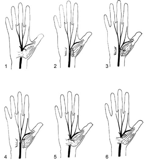
Figure 2 Anatomic variations of median nerve. 1. Usual branching of the median nerve; 2. Thenar branch leaving the median nerve within the carpal tunnel (subligamentous); 3. Transligamentous course of the thenar branch; 4. Thenar branch leaving median nerve at its ulnar aspect; 5. Thenar branch crosses over the top of the transverse carpal ligament; 6. Doubled thenar motor branch. Data from the article of Lanz (J Hand Surg Am. 1977;2:44-53).3)
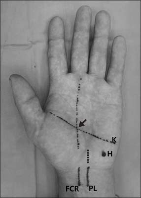
Figure 3 Kaplan cardinal line and anatomic landmarks. Dashed line is standard palmar incision and the solid arrow is the point that the recurrent motor branch of the median nerve usually enters the thenar muscles. K, Kaplan line; H, hamate hook; PL, palmaris longus; FCR, flexor carpi radialis.

Figure 4 Katz and Stirrat hand diagram.19) (A) Classic pattern. Symptoms affect at least two of digits 1, 2, or 3. The classic pattern permits symptoms in the fourth and fifth digits, wrist pain, and radiation of pain proximal to the wrist, but it does not allow symptoms on the palm or dorsum of the hand. (B) Probable pattern. Same symptom pattern as classic, except palmar symptoms are allowed unless confined solely to the ulnar aspect. In the possible pattern (not shown), symptoms involve only one of digits 1, 2, or 3. (C) Unlikely pattern. No symptoms are present in digits 1, 2, or 3.
Reference
-
1. Rotman MB, Donovan JP. Practical anatomy of the carpal tunnel. Hand Clin. 2002; 18:219–230.
Article2. Szabo RM, Pettey J. Bilateral median nerve bifurcation with an accessory compartment within the carpal tunnel. J Hand Surg Br. 1994; 19:22–23.
Article3. Lanz U. Anatomical variations of the median nerve in the carpal tunnel. J Hand Surg Am. 1977; 2:44–53.
Article4. Mackinnon SE, Dellon AL. Anatomic investigations of nerves at the wrist: I. Orientation of the motor fascicle of the median nerve in the carpal tunnel. Ann Plast Surg. 1988; 21:32–35.
Article5. Davlin LB, Aulicino PL, Bergfield TL. Anatomical variations of the median nerve at the wrist. Orthop Rev. 1992; 21:955–959.6. Riordan D, Kaplan E. Surface anatomy of the hand and wrist. In : Spinner M, editor. Kaplan's functional and surgical anatomy of the hand. Philadelphia: Lippincott;1984. p. 353–357.7. Taleisnik J. The palmar cutaneous branch of the median nerve and the approach to the carpal tunnel. An anatomical study. J Bone Joint Surg Am. 1973; 55:1212–1217.8. May JW Jr, Rosen H. Division of the sensory ramus communicans between the ulnar and median nerves: a complication following carpal tunnel release. A case report. J Bone Joint Surg Am. 1981; 63:836–838.9. Linburg RM, Comstock BE. Anomalous tendon slips from the flexor pollicis longus to the flexor digitorum profundus. J Hand Surg Am. 1979; 4:79–83.10. Szabo RM. Acute carpal tunnel syndrome. Hand Clin. 1998; 14:419–429.
Article11. Michelsen H, Posner MA. Medical history of carpal tunnel syndrome. Hand Clin. 2002; 18:257–268.
Article12. Ekman-Ordeberg G, Sälgeback S, Ordeberg G. Carpal tunnel syndrome in pregnancy. A prospective study. Acta Obstet Gynecol Scand. 1987; 66:233–235.
Article13. Strömberg T, Dahlin LB, Lundborg G. Hand problems in 100 vibration-exposed symptomatic male workers. J Hand Surg Br. 1996; 21:315–319.
Article14. Dias JJ, Burke FD, Wildin CJ, Heras-Palou C, Bradley MJ. Carpal tunnel syndrome and work. J Hand Surg Br. 2004; 29:329–333.
Article15. Massy-Westropp N, Grimmer K, Bain G. A systematic review of the clinical diagnostic tests for carpal tunnel syndrome. J Hand Surg Am. 2000; 25:120–127.
Article16. Graham B, Regehr G, Naglie G, Wright JG. Development and validation of diagnostic criteria for carpal tunnel syndrome. J Hand Surg Am. 2006; 31:919–924.
Article17. MacDermid JC, Wessel J. Clinical diagnosis of carpal tunnel syndrome: a systematic review. J Hand Ther. 2004; 17:309–319.
Article18. Durkan JA. A new diagnostic test for carpal tunnel syndrome. J Bone Joint Surg Am. 1991; 73:535–538.
Article19. Katz JN, Stirrat CR. A self-administered hand diagram for the diagnosis of carpal tunnel syndrome. J Hand Surg Am. 1990; 15:360–363.
Article20. Jablecki CK, Andary MT, So YT, Wilkins DE, Williams FH. AAEM Quality Assurance Committee. Literature review of the usefulness of nerve conduction studies and electromyography for the evaluation of patients with carpal tunnel syndrome. Muscle Nerve. 1993; 16:1392–1414.
Article21. Graham B. The value added by electrodiagnostic testing in the diagnosis of carpal tunnel syndrome. J Bone Joint Surg Am. 2008; 90:2587–2593.
Article22. El Miedany YM, Aty SA, Ashour S. Ultrasonography versus nerve conduction study in patients with carpal tunnel syndrome: substantive or complementary tests? Rheumatology (Oxford). 2004; 43:887–895.
Article23. Hobson-Webb LD, Massey JM, Juel VC, Sanders DB. The ultrasonographic wrist-to-forearm median nerve area ratio in carpal tunnel syndrome. Clin Neurophysiol. 2008; 119:1353–1357.
Article24. Mallouhi A, Pülzl P, Trieb T, Piza H, Bodner G. Predictors of carpal tunnel syndrome: accuracy of gray-scale and color Doppler sonography. AJR Am J Roentgenol. 2006; 186:1240–1245.
Article25. Nakamichi K, Tachibana S. Restricted motion of the median nerve in carpal tunnel syndrome. J Hand Surg Br. 1995; 20:460–464.
Article26. Fowler JR, Gaughan JP, Ilyas AM. The sensitivity and specificity of ultrasound for the diagnosis of carpal tunnel syndrome: a meta-analysis. Clin Orthop Relat Res. 2011; 469:1089–1094.
Article27. Burke DT, Burke MM, Stewart GW, Cambré A. Splinting for carpal tunnel syndrome: in search of the optimal angle. Arch Phys Med Rehabil. 1994; 75:1241–1244.
Article28. Manente G, Torrieri F, Di Blasio F, Staniscia T, Romano F, Uncini A. An innovative hand brace for carpal tunnel syndrome: a randomized controlled trial. Muscle Nerve. 2001; 24:1020–1025.
Article29. Chang MH, Chiang HT, Lee SS, Ger LP, Lo YK. Oral drug of choice in carpal tunnel syndrome. Neurology. 1998; 51:390–393.
Article30. Celiker R, Arslan S, Inanici F. Corticosteroid injection vs. nonsteroidal antiinflammatory drug and splinting in carpal tunnel syndrome. Am J Phys Med Rehabil. 2002; 81:182–186.31. Ellis J, Folkers K, Watanabe T, et al. Clinical results of a cross-over treatment with pyridoxine and placebo of the carpal tunnel syndrome. Am J Clin Nutr. 1979; 32:2040–2046.
Article32. Spooner GR, Desai HB, Angel JF, Reeder BA, Donat JR. Using pyridoxine to treat carpal tunnel syndrome. Randomized control trial. Can Fam Physician. 1993; 39:2122–2127.33. Kasdan ML, Janes C. Carpal tunnel syndrome and vitamin B6. Plast Reconstr Surg. 1987; 79:456–462.
Article34. Jacobson MD, Plancher KD, Kleinman WB. Vitamin B6 (pyridoxine) therapy for carpal tunnel syndrome. Hand Clin. 1996; 12:253–257.
Article35. Edgell SE, McCabe SJ, Breidenbach WC, LaJoie AS, Abell TD. Predicting the outcome of carpal tunnel release. J Hand Surg Am. 2003; 28:255–261.
Article36. Gelberman RH, Aronson D, Weisman MH. Carpal-tunnel syndrome. Results of a prospective trial of steroid injection and splinting. J Bone Joint Surg Am. 1980; 62:1181–1184.
Article37. Herskovitz S, Berger AR, Lipton RB. Low-dose, short-term oral prednisone in the treatment of carpal tunnel syndrome. Neurology. 1995; 45:1923–1925.
Article38. Chang MH, Ger LP, Hsieh PF, Huang SY. A randomised clinical trial of oral steroids in the treatment of carpal tunnel syndrome: a long term follow up. J Neurol Neurosurg Psychiatry. 2002; 73:710–714.
Article39. Aulisa L, Tamburrelli F, Padua R, Romanini E, Lo Monaco M, Padua L. Carpal tunnel syndrome: indication for surgical treatment based on electrophysiologic study. J Hand Surg Am. 1998; 23:687–691.
Article40. Hui AC, Wong S, Leung CH, et al. A randomized controlled trial of surgery vs steroid injection for carpal tunnel syndrome. Neurology. 2005; 64:2074–2078.
Article41. Agee JM, McCarroll HR Jr, Tortosa RD, Berry DA, Szabo RM, Peimer CA. Endoscopic release of the carpal tunnel: a randomized prospective multicenter study. J Hand Surg Am. 1992; 17:987–995.
Article42. Chow JC. Endoscopic release of the carpal ligament: a new technique for carpal tunnel syndrome. Arthroscopy. 1989; 5:19–24.
Article43. Thoma A, Veltri K, Haines T, Duku E. A systematic review of reviews comparing the effectiveness of endoscopic and open carpal tunnel decompression. Plast Reconstr Surg. 2004; 113:1184–1191.
Article44. Macdermid JC, Richards RS, Roth JH, Ross DC, King GJ. Endoscopic versus open carpal tunnel release: a randomized trial. J Hand Surg Am. 2003; 28:475–480.
Article45. Nagle DJ. Endoscopic carpal tunnel release. Hand Clin. 2002; 18:307–313.
Article46. Cook AC, Szabo RM, Birkholz SW, King EF. Early mobilization following carpal tunnel release. A prospective randomized study. J Hand Surg Br. 1995; 20:228–230.47. Mondelli M, Reale F, Sicurelli F, Padua L. Relationship between the self-administered Boston questionnaire and electrophysiological findings in follow-up of surgically-treated carpal tunnel syndrome. J Hand Surg Br. 2000; 25:128–134.
Article48. Gellman H, Kan D, Gee V, Kuschner SH, Botte MJ. Analysis of pinch and grip strength after carpal tunnel release. J Hand Surg Am. 1989; 14:863–864.
Article49. Ludlow KS, Merla JL, Cox JA, Hurst LN. Pillar pain as a postoperative complication of carpal tunnel release: a review of the literature. J Hand Ther. 1997; 10:277–282.50. Kim JK, Kim YK. Predictors of scar pain after open carpal tunnel release. J Hand Surg Am. 2011; 36:1042–1046.
Article51. Ring D, Kadzielski J, Fabian L, Zurakowski D, Malhotra LR, Jupiter JB. Self-reported upper extremity health status correlates with depression. J Bone Joint Surg Am. 2006; 88:1983–1988.
Article52. Amadio PC. Interventions for recurrent/persistent carpal tunnel syndrome after carpal tunnel release. J Hand Surg Am. 2009; 34:1320–1322.
Article53. Stütz N, Gohritz A, van Schoonhoven J, Lanz U. Revision surgery after carpal tunnel release--analysis of the pathology in 200 cases during a 2 year period. J Hand Surg Br. 2006; 31:68–71.54. Steyers CM. Recurrent carpal tunnel syndrome. Hand Clin. 2002; 18:339–345.
Article
- Full Text Links
-
- Actions
-
Cited
- CITED
-
- Close
- Share
-
- Similar articles
-
- Ultrasound-Guided Nerve Hydrodissection for Carpal Tunnel Syndrome
- Carpal Tunnel Syndrome in Children with Hypogammaglobulinemia: Case Report
- The Current Concepts for the Pathophysiology of Idiopathic Carpal Tunnel Syndrome
- The Carpal compression Test for Diagnosing Carpal Tunnel Syndrome
- Usefulness of Ultrasound for Carpal Tunnel Syndrome Proven in Meta-Analysis Studies
