The Diagnostic Strategy for Malignant Bone Tumors
- Affiliations
-
- 1Department of Orthopaedic Surgery, Yeungnam University Medical Center, Daegu, Korea. chpark77@naver.com
- KMID: 2185083
- DOI: http://doi.org/10.4055/jkoa.2015.50.6.429
Abstract
- Malignant bone tumors would be classified as primary malignant bone tumors, secondary malignant bone tumors, and metastatic bone tumors. Primary malignant bone tumors are rare diseases occupying 1% of adult cancers, and 6% of pediatric cancers. The chief complaint of malignant bone tumor patients is pain different from that of malignant soft tissue tumor patients. Diagnostic procedures start with clinical evaluation including current illness, past medical history, family history, and physical examination. Then we take a radiograph first and obtain important and diagnostic clues from it. However pathological diagnosis and information about the extent of tumor are required to obtain a more definite diagnosis and staging. Examinations for detection of local and systemic tumor extent are scintigraphy, computed tomography (CT), magnetic resonance imaging, and positron emission tomography-CT. If the clinical and radiographic information suggests aggressive or malignant bone tumor, the patient should be referred to a bone tumor specialist without further evaluations.
Keyword
MeSH Terms
Figure
-
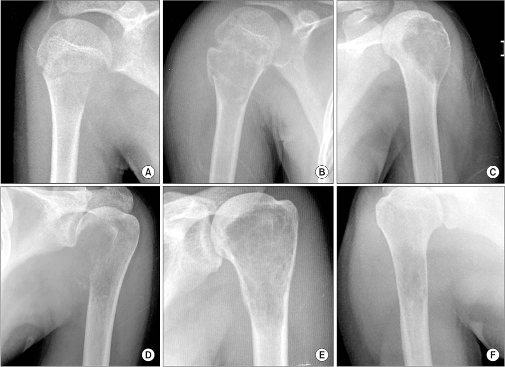
Figure 1 Various bone tumors in the proximal humerus according to age. (A) Langerhans cell histiocytosis in a 7-year-old patient. (B) Simple bone cyst in a 12-year-old patient. (C) Chondroblastoma in an 18-year-old patient. (D) Osteosarcoma in a 19-year-old patient. (E) Giant cell tumor in a 27-yearold patient. (F) Metastasis of lung cancer in a 51-year-old patient.
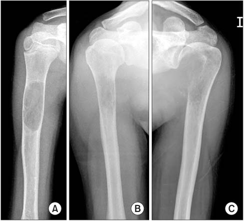
Figure 2 Patterns of bone destruction. (A) Geographic. (B) Moth-eaten. (C) Permeative.

Figure 3 Periosteal reactions of malignant bone tumors. (A, B) Onion peel appearance. (C, D) Sunburst appearance with Codman's triangle.
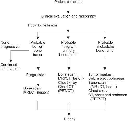
Figure 4 Diagnostic strategy for a bone tumor. MRI, magnetic resonance imaging; CT, computed tomography; PET/CT, positron emission tomography-CT.

Figure 5 Multiple bone metastases were detected on the whole body bone scan (Tc-99m-MDP scan).
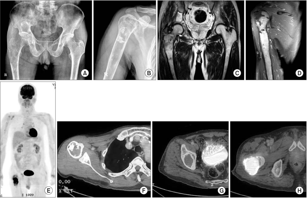
Figure 6 The effectiveness of positron emission tomography-computed tomography (PET-CT) in differentiation of benign and malignant lesions. (A, B) A 65-year-old man came to the hospital with right hip pain and showed 3 bone lesions (right supra acetabulum, right proximal femur, right humerus) in a radiograph suggesting multiple fibrous dysplasia. (C, D) There were no characteristic findings on magnetic resonance imaging to differentiate malignant change among three lesions. (E-H) PET-CT differentiated malignant change of the right proximal femoral lesion (high fluorodeoxyglucose [FDG, 18F] uptake) from others (right supra acetabulum and right humerus).
Reference
-
1. Souhami R. Incidence and aetiology of malignant primary bone tumours. Bailliere's Clin Oncol. 1987; 1:1–20.2. Jemal A, Murray T, Samuels A, Ghafoor A, Ward E, Thun MJ. Cancer statistics, 2003. CA Cancer J Clin. 2003; 53:5–26.3. HaDuong JH, Martin AA, Skapek SX, Mascarenhas L. Sarcomas. Pediatr Clin North Am. 2015; 62:179–200.4. Simon MA, Springfield DS, Conrad EU. Surgery for bone and soft-tissue tumors. Philadelphia: Lippincott-Raven;1998.5. Grimer RJ, Briggs TW. Earlier diagnosis of bone and soft-tissue tumours. J Bone Joint Surg Br. 2010; 92:1489–1492.6. Temple HT, Scully SP, Aboulafia AJ. Benign bone tumors. Instr Course Lect. 2002; 51:429–439.7. Moore DD, Luu HH. Osteosarcoma. Cancer Treat Res. 2014; 162:65–92.8. Weber K, Damron TA, Frassica FJ, Sim FH. Malignant bone tumors. Instr Course Lect. 2008; 57:673–688.9. Scully SP, Ghert MA, Zurakowski D, Thompson RC, Gebhardt MC. Pathologic fracture in osteosarcoma: prognostic importance and treatment implications. J Bone Joint Surg Am. 2002; 84:49–57.10. Unni KK, Inwards CY, Bridge JA, Kindblum LG, Wold LE. Tumours of bones and joints. Atlas of tumour pathology 4th series. 1st ed. Washington, DC: Armed Forces Institute of Pathology;2005.11. Lodwick GS, Wilson AJ, Farrell C, Virtama P, Dittrich F. Determining growth rates of focal lesions of bone from radiographs. Radiology. 1980; 134:577–583.12. Teo HE, Peh WC. The role of imaging in the staging and treatment planning of primary malignant bone tumors in children. Eur Radiol. 2004; 14:465–475.13. Vlychou M, Athanasou NA. Radiological and pathological diagnosis of paediatric bone tumours and tumour-like lesions. Pathology. 2008; 40:196–216.14. Shimose S, Kubo T, Fujimori J, Furuta T, Ochi M. A novel assessment method of serum alkaline phosphatase for the diagnosis of osteosarcoma in children and adolescents. J Orthop Sci. 2014; 19:997–1003.15. Mavrogenis AF, Angelini A, Vottis C, et al. State-of-the-art approach for bone sarcomas. Eur J Orthop Surg Traumatol. 2015; 25:5–15.16. Mankin HJ, Lange TA, Spanier SS. The hazards of biopsy in patients with malignant primary bone and soft-tissue tumors. J Bone Joint Surg Am. 1982; 64:1121–1127.17. Mentzel HJ, Kentouche K, Sauner D, et al. Comparison of whole-body STIR-MRI and 99mTc-methylene-diphosphonate scintigraphy in children with suspected multifocal bone lesions. Eur Radiol. 2004; 14:2297–2302.18. Shin DS, Cho IH. Predicting the response of preoperative chemotherapy in osteosarcoma by thallium-201 scintigraphy. J Korean Orthop Assoc. 2003; 38:722–727.19. Fenoy AJ, Greenlee JD, Menezes AH, et al. Primary bone tumors of the spine in children. J Neurosurg. 2006; 105:4 Suppl. 252–260.20. Yu H, Cui JL, Cui SJ, Sun YC, Cui FZ. Differentiating benign from malignant bone tumors using fluid-fluid level features on magnetic resonance imaging. Korean J Radiol. 2014; 15:757–763.21. Shin DS, Shon OJ, Han DS, Choi JH, Chun KA, Cho IH. The clinical efficacy of (18)F-FDG-PET/CT in benign and malignant musculoskeletal tumors. Ann Nucl Med. 2008; 22:603–609.22. Yang HL, Liu T, Wang XM, Xu Y, Deng SM. Diagnosis of bone metastases: a meta-analysis comparing 18FDG PET, CT, MRI and bone scintigraphy. Eur Radiol. 2011; 21:2604–2617.23. Shin DS, Shon OJ, Byun SJ, Choi JH, Chun KA, Cho IH. Differentiation between malignant and benign pathologic fractures with F-18-fluoro-2-deoxy-D-glucose positron emission tomography/computed tomography. Skeletal Radiol. 2008; 37:415–421.24. Maciel MJ, Tyng CJ, Barbosa PN, et al. Computed tomography-guided percutaneous biopsy of bone lesions: rate of diagnostic success and complications. Radiol Bras. 2014; 47:269–274.25. Heindel W, Gübitz R, Vieth V, Weckesser M, Schober O, Schäfers M. The diagnostic imaging of bone metastases. Dtsch Arztebl Int. 2014; 111:741–747.26. Rybak LD, Rosenthal DI. Radiological imaging for the diagnosis of bone metastases. Q J Nucl Med. 2001; 45:53–64.
