MDCT Application of Thoracic Imaging
- Affiliations
-
- 1Department of Radiology and Imaging Science, Sungkyunkwan University School of Medicine, Korea. chungmj@smc.samsung.co.kr
- KMID: 2184795
- DOI: http://doi.org/10.5124/jkma.2007.50.1.57
Abstract
- Multidetector-row computed tomography (MDCT) provides new opportunities and poses challenges for medical imaging to radiologists and physicians. Isotropic imaging (similar resolution in three dimensional directions) allows in-depth views of anatomy and disease. Ultra-fast scan enables whole-body volume imaging within a single breath hold and thus the reduction of contrast medium consumption. CT volume data sets can be used for threedimensional visualization of the whole body, with which the detailed and comprehensive interpretation of thoracic anatomy and specific disease location and extent is plausible. Moreover, four-dimensional CT imaging can be possible and therefore, we can observe and quantify cardiopulmonary functions without invasive procedures. The author reviews briefly the application of MDCT for the thoracic imaging.
MeSH Terms
Figure
-
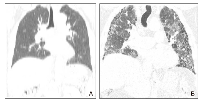
Figure 1 Comparison of multiplanar image reformatted from conventional CT and MDCT (A) Coronal reformatted image from single spiral CT with 5mm thick axial image (B) Coronal reformatted image from sixteen multidetector-row CT with 1.25mm thick axial image
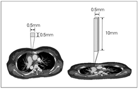
Figure 2 Comparison of pixel and voxel. One pixel of axial CT has 0.5 × 0.5 mm size on XY plan. However Z axis length of this pixel on three dimensions is long to 10mm. This voxel shows unusually long hexahedron
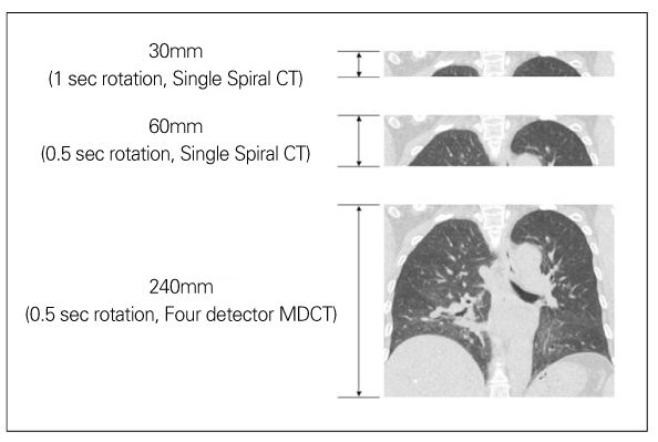
Figure 3 How long scan with 1 mm slices thickness in 30 second scan time?
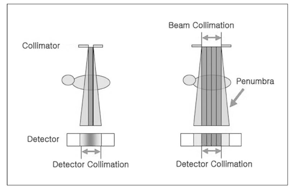
Figure 4 Diagram of penumbra effect
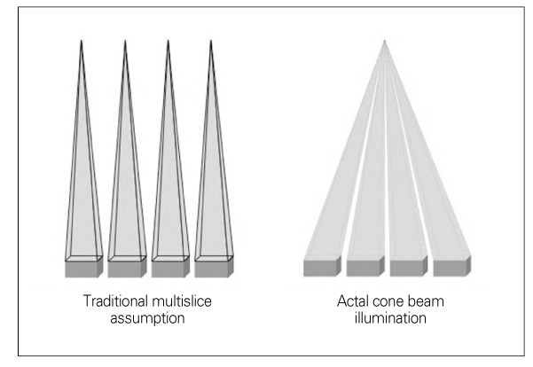
Figure 5 Diagram of beam transmission with traditional axial scan vs. cone beam scan of MDCT
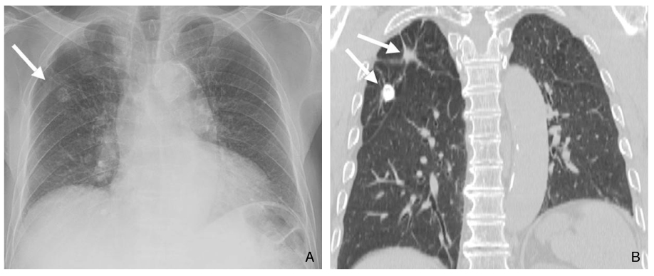
Figure 6 Comprehensive interpretation of anatomy using multiplanar image (A) Plain radiography shows calcified granuloma in right upper lung zone (arrow) (B) Coronal reformatted image of MDCT shows similar view with (A) It shows calcified granuloma also and fibrotic bands in right upper lobe as an additional finding (arrows)
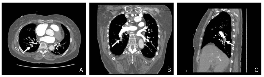
Figure 7 Pulmonary thromboembolism on multplanar CT angiography. Filling defect (arrow) in right inferior pulmonary artery is conspicuous within highly enhanced vascular structure (A) Axial image (B) Coronal image (C) Sagital image
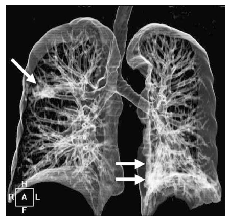
Figure 8 Virtual bronchography using three dimensional volume rendering technique. Tubular structures with high (virtual) enhancement in right upper lobe and left lower lobe (arrows) are tubular bronchiectasis
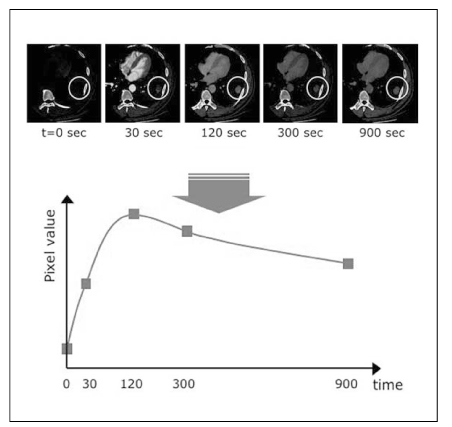
Figure 9 Enhancement dynamic CT of pulmonary nodule. Serial images obtained at 30-second intervals and at similar levels show enhancement dynamics of nodule. We can extract objective attenuation of nodule and plot time-attenuation curve
Reference
-
1. Taguchi K, Aradate H. Algorithm for image reconstruction in multi-slice helical CT. Med Phys. 1998. 25:550–561.
Article2. De Smedt E, Potvliege R, Pimontel-Appel B, Claus E, Vignaud J. High resolution CT-scan of the temporal bone; a preliminary report. J Belge Radiol. 1980. 63:205–212.3. Todo G, Herman PG. High-resolution computed tomography of the pig lung. Invest Radiol. 1986. 21:689–696.
Article4. Kohler T, Proksa R, Bontus C, Grass M, Timmer J. Artifact analysis of approximate helical cone-beam CT reconstruction algorithms. Med Phys. 2002. 29:51–64.
Article5. Zou Y, Sidky EY, Pan X. Partial volume and aliasing artefacts in helical cone-beam CT. Phys Med Biol. 2004. 49:65–75.
Article6. Yang K, Kwan AL, Miller DF, Boone JM. A geometric calibration method for cone beam CT systems. Med Phys. 2006. 33:1695–1706.
Article7. Mori S, Endo M, Kondo C, Tanada S. Physical evaluation of the weighted Feldkamp algorithms applied to the 256-detector row CT scanner for volumetric cine imaging. Acad Radiol. 2006. 13:701–712.
Article8. Schmidt TG, Fahrig R, Pelc NJ. A three-dimensional reconstruction algorithm for an inverse-geometry volumetric CT system. Med Phys. 2005. 32:3234–3245.
Article9. Remy J, Remy-Jardin M, Artaud D, Fribourg M. Multiplanar and three-dimensional reconstruction techniques in CT: impact on chest diseases. Eur Radiol. 1998. 8:335–351.
Article10. Studler U, Gluecker T, Bongartz G, Roth J, Steinbrich W. Image quality from high-resolution CT of the lung: comparison of axial scans and of sections reconstructed from volumetric data acquired using MDCT. AJR Am J Roentgenol. 2005. 185:602–607.
Article11. Sung YM, Lee KS, Yi CA, Yoon YC, Kim TS, Kim S. Additional coronal images using low-milliamperage multidetector-row computed tomography: effectiveness in the diagnosis of bronchiectasis. J Comput Assist Tomogr. 2003. 27:490–495.
Article12. Chung MJ, Lee JH, Lee KS, Yoon YC, Kwon OJ, Kim TS. Bronchial and nonbronchial systemic arteries in patients with hemoptysis: depiction on MDCT angiography. AJR Am J Roentgenol. 2006. 186:649–655.
Article13. Yoon YC, Lee KS, Jeong YJ, Shin SW, Chung MJ, Kwon OJ. Hemoptysis: bronchial and nonbronchial systemic arteries at 16-detector row CT. Radiology. 2005. 234:292–298.
Article14. Carraro M, Malalan F, Antonione R, Stacul F, Cova M, Petz S, Assante M, Grynne B, Haider T, Palma LD, Faccini L. Effects of a dimeric vs a monomeric nonionic contrast medium on renal function in patients with mild to moderate renal insufficiency: a double-blind, randomized clinical trial. Eur Radiol. 1998. 8:144–147.
Article15. Spataro RF, Fischer HW, Kormano M. Clinical comparison of Hexabrix, iopamidol, and urografin-60 in whole body computed tomography. Invest Radiol. 1984. 19:372–375.
Article16. Donadio C, Tramonti G, Lucchesi A, Giordani R, Lucchetti A, Bianchi C. Tubular toxicity is the main renal effect of contrast media. Ren Fail. 1996. 18:647–656.
Article17. Bae KT. Peak contrast enhancement in CT and MR angiography: when does it occur and why? Pharmacokinetic study in a porcine model. Radiology. 2003. 227:809–816.
Article18. Bae KT, Heiken JP, Brink JA. Aortic and hepatic peak enhancement at CT: effect of contrast medium injection rate--pharmacokinetic analysis and experimental porcine model. Radiology. 1998. 206:455–464.
Article19. Jeong YJ, Lee KS, Yoon YC, Kim TS, Chung MJ, Kim S. Evaluation of small pulmonary arteries by 16-slice multidetector computed tomography: Optimum slab thickness in condensing transaxial images converted into maximum intensity projection images. J Comput Assist Tomogr. 2004. 28:195–203.
Article20. Prologo JD, Gilkeson RC, Diaz M, Cummings M. The effect of single-detector CT versus MDCT on clinical outcomes in patients with suspected acute pulmonary embolism and negative results on CT pulmonary angiography. AJR Am J Roentgenol. 2005. 184:1231–1235.
Article21. Perrier A, Roy PM, Sanchez O, Le Gal G, Meyer G, Gourdier AL, Furber A, Revel MP, Howarth N, Davido A, Bounameaux H. Multidetector-row computed tomography in suspected pulmonary embolism. N Engl J Med. 2005. 04. 28. 352:1760–1768.
Article22. Lee KS, Yoon JH, Kim TK, Kim JS, Chung MP, Kwon OJ. Evaluation of tracheobronchial disease with helical CT with multiplanar and three-dimensional reconstruction: correlation with bronchoscopy. Radiographics. 1997. 17:555–567. discussion 568 - 570.
Article23. Yi CA, Lee KS, Kim EA. Solitary pulmonary nodules: dynamic enhanced multi-detector row CT study and comparison with vascular endothelial growth factor and microvessel density. Radiology. 2004. 233:191–199.
Article24. Jeong YJ, Lee KS, Jeong SY. Solitary pulmonary nodule: characterization with combined wash-in and washout features at dynamic multi-detector row CT. Radiology. 2005. 237:675–683.
Article25. Chung MJ, Lee KS, Han J, Sung YM, Chong S, Kwon OJ. Pulmonary sclerosing hemangioma presenting as solitary pulmonary nodule: dynamic CT findings and histopathologic comparisons. AJR Am J Roentgenol. 2006. 187:430–437.
Article26. Naidich DP, Gruden JF, McGuinness G, McCauley DI, Bhalla M. Volumetric (helical/spiral) CT (VCT) of the airways. J Thorac Imaging. 1997. 12:11–28.
Article27. Touliopoulos P, Costello P. Helical (spiral) CT of the thorax. Radiol Clin North Am. 1995. 33:843–861.28. Schueller-Weidekamm C, Wassermann E, Redl H, Prokop M, Zimpfer M, Herold C, Germann P, Ullrich R. Dynamic CT measurement of pulmonary enhancement in piglets with experimental acute respiratory distress syndrome. Radiology. 2006. 239:398–405.
Article29. Hoffman EA, Chon D. Computed tomography studies of lung ventilation and perfusion. Proc Am Thorac Soc. 2005. 2:492–498.
Article30. Guerrero T, Sanders K, Castillo E, Zhang Y, Bidaut L, Pan T, Komaki R. Dynamic ventilation imaging from four-dimensional computed tomography. Phys Med Biol. 2006. 51:777–791.
Article31. Lee KS, Ernst A, Trentham DE, Lunn W, Feller-Kopman DJ, Boiselle PM. Relapsing polychondritis: prevalence of expiratory CT airway abnormalities. Radiology. 2006. 240:565–573.
Article
