Ischemic Neuropathy Associated with Livedoid Vasculitis
- Affiliations
-
- 1Department of Neurology, Seoul Medical Center, Seoul, Korea.
- 2Department of Neurology, Seoul National University Hospital, Seoul, Korea. jjsaint@snu.ac.kr
- 3Department of Neurology, Medical University of South Carolina, Charleston, SC, USA.
- 4Department of Neurology, Seoul Boramae Hospital, Seoul, Korea.
- 5Department of Neurology, Seoul National University Bundang Hospital, Seoul, Korea.
- KMID: 2179011
- DOI: http://doi.org/10.3988/jcn.2011.7.4.233
Abstract
- BACKGROUND
Livedoid vasculitis is a chronic dermatological problem with an unclear etiology. Clinical findings are petechiae with painful ulcers in both lower extremities, which heal to become hyperpigmented and porcelain-white satellite lesions. There are only a few reported cases of livedoid vasculitis presenting in combination with peripheral neuropathy.
CASE REPORT
We report the first case of a Korean patient presenting with mononeuritis multiplex combined with livedoid vasculitis, which was confirmed by electrophysiological and pathological studies.
CONCLUSIONS
Our report supports the possible vaso-occlusive etiology of livedoid vasculitis in multifocal ischemic neuropathy.
Keyword
Figure
-
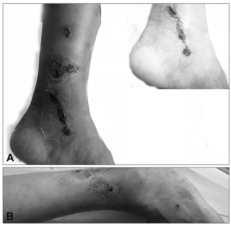
Fig. 1 Multiple painful ulcerations with healed white scarring in the bilateral lateral malleolar area. A: left lateral malleolar area. B: right lateral malleolar area.

Fig. 2 Histopathology findings of a sural nerve biopsy. A: Extensive infarct of the peripheral nerve and Schwann cells (hematoxylin-eosin stain, original magnification ×40). B: Endoneurial capillary ectasia and congestion with hemorrhage (hematoxylin-eosin stain, original magnification ×200). C: Intravascular thrombosis with mild lymphocytic infiltration present in the epineurium. There was no definite neutrophilia or leukocytoclasia, suggesting vasculitis (i: hematoxylin-eosin stain, ii: leukocyte common antigen stain, original magnification ×200). D: Electron micrograph showing marked degeneration of axons and their myelin sheath, with the Schwann cell cytoplasm containing many autophagic vacuoles and myelin figures.
Cited by 1 articles
-
Successful Treatment of Livedoid Vasculitis with Primary Antiphospholipid Syndrome by Using Aspirin and Low Dose Warfarin Combination Therapy
Byoung Joon So, Jae Beom Park, Min Gun Yoo, Il-Hwan Kim, Sang Wook Son
Ann Dermatol. 2015;27(5):614-615. doi: 10.5021/ad.2015.27.5.614.
Reference
-
1. Bard JW, Winkelmann RK. Livedo vasculitis. Segmental hyalinizing vasculitis of the dermis. Arch Dermatol. 1967. 96:489–499.
Article2. Papi M, Diodona B, De Pità O, Silvestri L, Ferranti G, Gantcheva M, et al. PURPLE (atrophie blanche): clinical, histological and immunological study of twelve patients. J Eur Acad Dermatol Venereol. 1997. 9:129–133.
Article3. Winkelmann RK, Schroeter AL, Kierland RR, Ryan TM. Clinical studies of livedoid vasculitis: (segmental hyalinizing vasculitis). Mayo Clin Proc. 1974. 49:746–750.4. Toth C, Trotter M, Clark A, Zochodne D. Mononeuropathy multiplex in association with livedoid vasculitis. Muscle Nerve. 2003. 28:634–639.
Article5. Osada S, Kimura Y, Kawana S. Case of livedoid vasculopathy with peripheral neuropathy successfully treated with low-dose warfarin. J Dermatol. 2010. 37:98–101.
Article6. Gibson LE, Su WP. Cutaneous vasculitis. Rheum Dis Clin North Am. 1995. 21:1097–1113.
Article7. Milstone LM, Braverman IM, Lucky P, Fleckman P. Classification and therapy of atrophie blanche. Arch Dermatol. 1983. 119:963–969.
Article8. Grattan CE, Burton JL, Boon AP. Sneddon's syndrome (livedo reticularis and cerebral thrombosis) with livedo vasculitis and anticardiolipin antibodies. Br J Dermatol. 1989. 120:441–447.
Article9. Grob JJ, Bonerandi JJ. Thrombotic skin disease as a marker of the anticardiolipin syndrome. Livedo vasculitis and distal gangrene associated with abnormal serum antiphospholipid activity. J Am Acad Dermatol. 1989. 20:1063–1069.10. Baccard M, Vignon-Pennamen MD, Janier M, Scrobohaci ML, Dubertret L. Livedo vasculitis with protein C system deficiency. Arch Dermatol. 1992. 128:1410–1411.
Article11. Pizzo SV, Murray JC, Gonias SL. Atrophie blanche. A disorder associated with defective release of tissue plasminogen activator. Arch Pathol Lab Med. 1986. 110:517–519.12. Tsutsui K, Shirasaki F, Takata M, Takehara K. Successful treatment of livedo vasculitis with beraprost sodium: a possible mechanism of thrombomodulin upregulation. Dermatology. 1996. 192:120–124.
Article13. Gibson GE, Li H, Pittelkow MR. Homocysteinemia and livedoid vasculitis. J Am Acad Dermatol. 1999. 40:279–281.
Article14. Calamia KT, Balabanova M, Perniciaro C, Walsh JS. Livedo (livedoid) vasculitis and the factor V Leiden mutation: additional evidence for abnormal coagulation. J Am Acad Dermatol. 2002. 46:133–137.
Article15. Khenifer S, Thomas L, Balme B, Dalle S. Livedoid vasculopathy: thrombotic or inflammatory disease? Clin Exp Dermatol. 2010. 35:693–698.
Article16. Jacobson RR, Krahenbuhl JL. Leprosy. Lancet. 1999. 353:655–660.
Article17. Ooi C, Dayan L. Syphilis. Diagnosis and management in general practice. Aust Fam Physician. 2002. 31:629–635.18. Jetton RL, Lazarus GS. Minidose heparin therapy for vasculitis of atrophie blanche. J Am Acad Dermatol. 1983. 8:23–26.
Article19. Klein KL, Pittelkow MR. Tissue plasminogen activator for treatment of livedoid vasculitis. Mayo Clin Proc. 1992. 67:923–933.
Article20. Drucker CR, Duncan WC. Antiplatelet therapy in atrophie blanche and livedo vasculitis. J Am Acad Dermatol. 1982. 7:359–363.
Article21. Dedichen J, Gjessing HC. Livedo reticularis with summer ulcerations; report of case treated with long-term anticoagulation therapy. Acta Med Scand Suppl. 1956. 319:74–78.22. Hsiao GH, Chiu HC. Livedoid vasculitis. Response to low-dose danazol. Arch Dermatol. 1996. 132:749–751.23. Rustin MH, Bunker CB, Dowd PM. Chronic leg ulceration with livedoid vasculitis, and response to oral ketanserin. Br J Dermatol. 1989. 120:101–105.
Article24. Hoogenberg K, Tupker RA, van Essen LH, Smit AJ, Kallenberg CG. Successful treatment of ulcerating livedo reticularis with infusions of prostacyclin. Br J Dermatol. 1992. 127:64–66.
Article25. Amital H, Levy Y, Shoenfeld Y. Use of intravenous immunoglobulin in livedo vasculitis. Clin Exp Rheumatol. 2000. 18:404–406.
- Full Text Links
-
- Actions
-
Cited
- CITED
-
- Close
- Share
-
- Similar articles
-
- Three Cases of Livedoid Vasculitis Improved by Low-dose Danazol
- A Case of Livedoid Vasculitis
- Ischemic Monomelic Neuropathy of Both Upper Extremity: A Complication of Hemodialysis Access Procedure
- Successful treatment of refractory livedoid vasculitis with rituximab and cyclophosphamide
- Livedoid Vasculitis: a Clinico-Pathological Study of 19 Patients
