Pneumocephalus in Patients With Orthostatic Headache
- Affiliations
-
- 1Department of Neurology, Eulji University College of Medicine, Daejeon, Korea. trumind@lycos.co.kr
- 2Department of Radiology, Eulji University College of Medicine, Daejeon, Korea.
- KMID: 2178928
- DOI: http://doi.org/10.3988/jcn.2008.4.2.89
Abstract
- Cerebrospinal fluid (CSF) leak or shunt overdrainage is a well-known cause of orthostatic headaches and low CSF pressures. We report two cases of orthostatic headache with pneumocephalus on brain imaging. The orthostatic headache developed after drainage of spinal operation site and epidural block. Brain MRI revealed characteristic findings of CSF hypovolemia including pachymeningeal enhancement and mild subdural fluid collections. Air was also observed in the ventricular or subarachnoid space in both patients, which might enter the subarachnoid or ventricular space during a procedure via the pressure gradient or an injection.
Figure
-
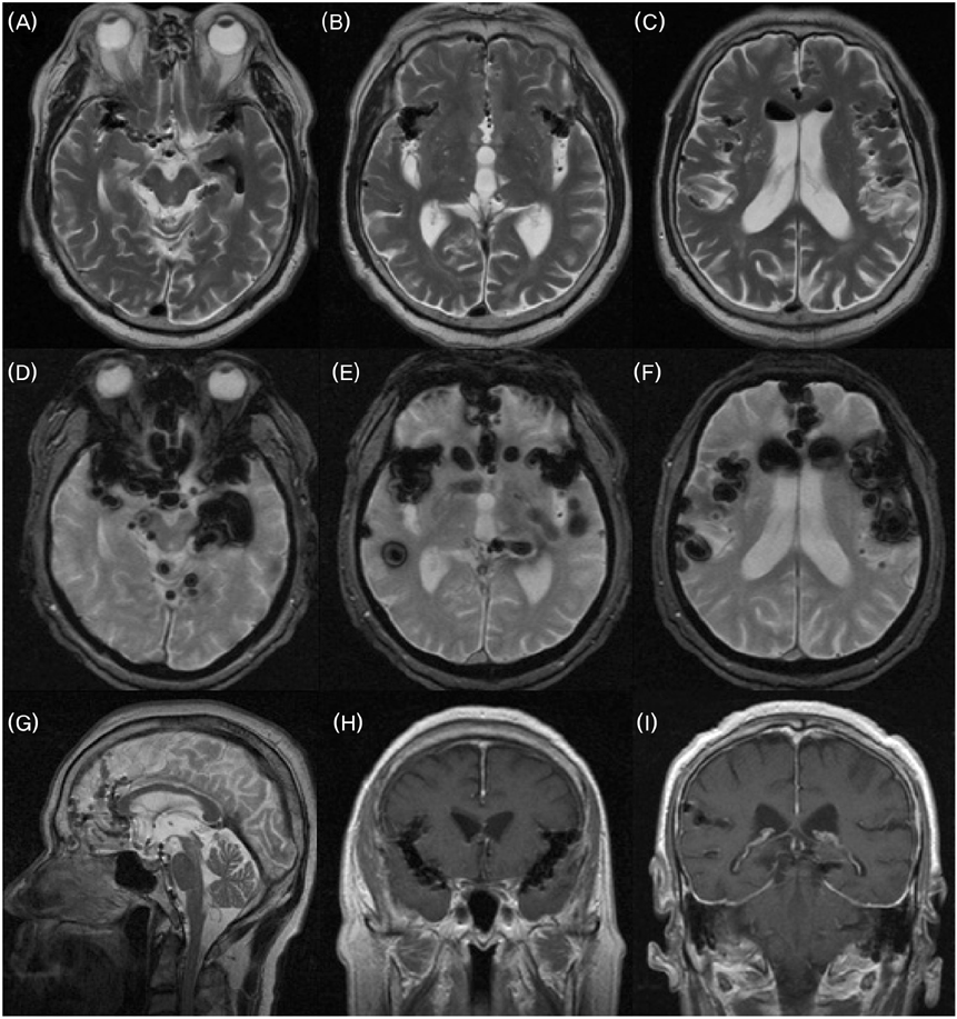
Figure 1 Brain MRI of case 1. T2-weighted axial images (A, B, and C) show massive pneumocephalus involving both lateral ventricles, basal cisterns, sylvian cisterns, the left ambient cistern, retrocerebellar cisterns, cerebellar folia, the anterior interhemispheric fissure, and cortical sulci (subarachnoid space). Gradient echo images (D, E, and F) show markedly exaggerated findings relative to T2-weighted axial images. Sagittal image (G) reveals the air pathway, and enhanced MRI coronal images (H and I) demonstrate diffuse nonnodular, uninterrupted pachymeningeal gadolinium enhancement.
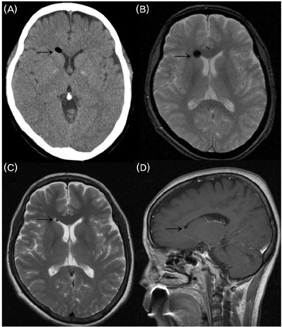
Figure 2 Brain images of case 2. Brain CT (A) was performed 1 day before brain MRI. Note the presence of exaggerated air in the right anterior frontal horn on the gradient echo image (B) relative to the brain CT and T2-weighted axial images (C). Slightly prominent extracerebral space at anterior frontal areas - which is suggestive of minimal subdural hygroma - is also observed on an enhanced T1-weighted sagittal MR image (D). Arrows indicate air in the right anterior frontal horn.
Cited by 1 articles
-
Pneumocephalus in a Patient with No Cerebrospinal Fluid Leakage after Lumbar Epidural Block - A Case Report -
Yeon Dong Kim, Jae Hun Lee, Yong Kwan Cheong
Korean J Pain. 2012;25(4):262-266. doi: 10.3344/kjp.2012.25.4.262.
Reference
-
1. Fishman RA, Dillon WP. Dural enhancement and cerebral displacement secondary to intracranial hypotension. Neurology. 1993. 43:609–611.
Article2. Mokri B. Headaches caused by decreased intracranial pressure: diagnosis and management. Curr Opin Neurol. 2003. 16:319–326.
Article3. Mokri B. The Monro-Kellie hypothesis: applications in CSF volume depletion. Neurology. 2001. 56:1746–1748.
Article4. Ozturk E, Kantarci M, Karaman K, Basekim CC, Kizilkaya E. Diffuse pneumocephalus associated with infratentorial and supratentorial hemorrhages as a complication of spinal surgery. Acta Radiol. 2006. 47:497–500.
Article5. Clevens RA, Marentette LJ, Esclamado RM, Wolf GT, Ross DA. Incidence and management of tension pneumocephalus after anterior craniofacial resection: case reports and review of the literature. Otolaryngol Head Neck Surg. 1999. 120:579–583.
Article6. Kozikowski GP, Cohen SP. Lumbar puncture associated with pneumocephalus: report of a case. Anesth Analg. 2004. 98:524–526.
Article7. Bilsky MH, Downey RJ, Kaplitt MG, Elowitz EH, Rusch VW. Tension pneumocephalus resulting from iatrogenic subarachnoid-pleural fistulae: report of three cases. Ann Thorac Surg. 2001. 71:455–457.
Article8. Chung SH, Lee SB, Kang MC, Yoon SS, Lee HJ, Chung KC. A case of spontaneous pneumocephalus associated with pneumococcal meningitis. J Korean Neurol Assoc. 2005. 23:425–427.9. Simopoulos T, Peeters-Asdourian C. Pneumocephalus after cervical epidural steroid injection. Anesth Analg. 2001. 92:1576–1577.
Article10. Kats JA, Lukin R, Bridenbaugh PO, Gunzenhauser L. Subdural intracranial air: an unusual cause of headache after epidural steroid injection. Anesthesiology. 1991. 74:615–618.11. Lucas DN, Kennedy A, Dob DP. Dural puncture and iatrogenic pneumocephalus with subsequent transverse myelitis in a parturient. Can J Anaesth. 2000. 47:1103–1106.
Article12. Lai TH, Fuh JL, Lirng FJ, Tsai PH, Wang SJ. Subdural haematoma in patients with spontaneous intracranial hypotension. Cephalagia. 2006. 27:133–138.
Article13. Han SR, Kim YJ, Km YI, Lee KS, Kim BS, Choo SW. A case of unexpected clinical course of spontaneous intracranial hypotension. J Korean Neurol Assoc. 1995. 13:129–132.
- Full Text Links
-
- Actions
-
Cited
- CITED
-
- Close
- Share
-
- Similar articles
-
- Pneumocephalus after an Epidural Injection
- Pneumocephalus after Epidural Steroid Injection: A case report
- Headache and Pneumocephalus after Lumbar Epidural Block: A case report
- Pneumocephalus after Interlaminar Lumbar Epidural Block
- Pneumocephalus after Inadvertent Dural Puncture during Epidural Block: A case report
