J Cardiovasc Ultrasound.
2014 Mar;22(1):46-47. 10.4250/jcu.2014.22.1.46.
Similar Morphology, but Different Function: Acute Improvement of Myocardial Longitudinal Strain after Percutaneous Transcatheter Aortic Valve Implantation Therapy in a Severe Aortic Stenosis Patient
- Affiliations
-
- 1Department of Cardiology, Internal Medicine, School of Medicine, Chungnam National University, Chungnam National University Hospital, Daejeon, Korea. jaehpark@cnu.ac.kr
- KMID: 2177458
- DOI: http://doi.org/10.4250/jcu.2014.22.1.46
Abstract
- No abstract available.
MeSH Terms
Figure
-
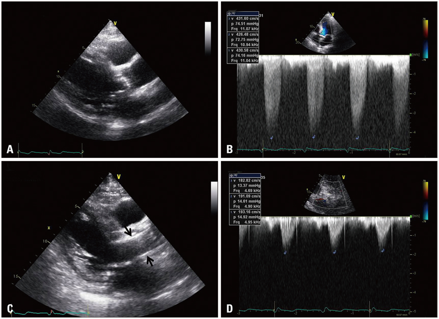
Fig. 1 The baseline parasternal long axis view demonstrates severe calcified aortic valve (A) with increased transaortic valve maximal velocity up to 4.3 m/sec (B). After the procedure, prosthetic valve is inserted in to the aortic valve area (C, arrows) and transvalvular maximal velocity is measured up to 1.9 m/sec (D).

Fig. 2 Bull's eye diagram shows clearly visible improvement in global longitudinal strain after the transfemoral aortic valve intervention [before (A), and one week after (B) the procedure].
Reference
-
1. Delgado V, Tops LF, van Bommel RJ, van der Kley F, Marsan NA, Klautz RJ, Versteegh MI, Holman ER, Schalij MJ, Bax JJ. Strain analysis in patients with severe aortic stenosis and preserved left ventricular ejection fraction undergoing surgical valve replacement. Eur Heart J. 2009; 30:3037–3047.
Article2. Cribier A, Eltchaninoff H, Bash A, Borenstein N, Tron C, Bauer F, Derumeaux G, Anselme F, Laborde F, Leon MB. Percutaneous transcatheter implantation of an aortic valve prosthesis for calcific aortic stenosis: first human case description. Circulation. 2002; 106:3006–3008.
Article
- Full Text Links
-
- Actions
-
Cited
- CITED
-
- Close
- Share
-
- Similar articles
-
- A Case of Severe Aortic Stenosis Patient With High Operative Risk Treated by Transcatheter Aortic-Valve Implantation
- Anesthetic considerations of percutaneous transcatheter aortic valve implantation: first attempt in Korea: A report of 2 cases
- Expanding transcatheter aortic valve replacement into uncharted indications
- Echocardiography in Transcatheter Aortic Valve Implantation and Mitral Valve Clip
- Contrast-free (Zero-contrast) TAVR for Severe Aortic Stenosis in Patient with Chronic Kidney Disease
