Evaluation of canal preparation with Ni-Ti rotary files by micro computed tomography
- Affiliations
-
- 1Department of Conservative Dentistry, College of Dentistry, Seoul National University, Korea. shbaek@snu.ac.kr
- KMID: 2175632
- DOI: http://doi.org/10.5395/JKACD.2004.29.4.378
Abstract
- The purpose of this study was to compare the effects of preparation with GT files and profiles .04 in shaping of root canals and reconstruct the three-dimensional root canal system using micro computed tomography. 40 canals of the extracted human mandibular molars were used, and randomly distributed into two experimental groups. In group 1, canals were prepared by GT files. In group 2, Profiles .04. were used. Apical preparation size was #30. For each tooth pre and post operative cross-sectional images were obtained by the micro CT at 50 micron intervals. Pre and post operative cross-sectional images of 1, 2, 3, 5, and 8mm from the apex were compared. For each section, canal area and centering ratio were determined. For each tooth pre- and post-operative root canal volume from the furcation to the apex of the roots was calculated by three-dimensional image software. Following results were obtained: 1. At 8mm from the apex, area of dentin removed by GT rotary file was significantly larger than that by Profile .04. And at the other levels there was not a significant difference. 2. There was a trend for GT rotary file to remain more centered in the canals than Profile .04 at all levels. But at 3mm level, there was a statistically significant difference. 3. In root canal volume increments after instrumentation, there was no significant difference between two groups.
Figure
-
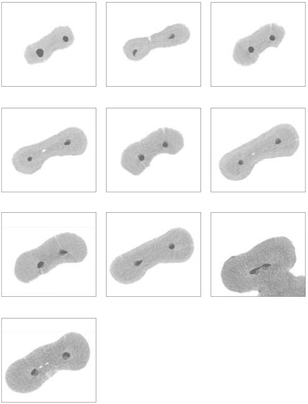
Figure 1 Examples of superimposed images (from the top to the bottom 1, 2, 3, 5 and 8 mm level respectively. left : GT file, right : Profile .04)

Figure 2 Reconstructed pulp space (before and after preparation of mesial canals)
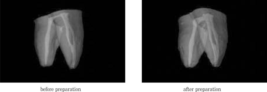
Figure 3 Reconstructed root canal system (before and after preparation of mesial canals
Cited by 2 articles
-
Comparative study on morphology of cross-section and cyclic fatigue test with different rotary NiTi files and handling methods
Jae-Gwan Kim, Kee-Yeon Kum, Eui-Seong Kim
J Korean Acad Conserv Dent. 2006;31(2):96-102. doi: 10.5395/JKACD.2006.31.2.096.Evaluation of apical canal shapes produced sequentially during instrumentation with stainless steel hand and Ni-Ti rotary instruments using Micro-computed tomography
Woo-Jin Lee, Jeong-Ho Lee, Kyung-A Chun, Min-Seock Seo, Yeon-Jee Yoo, Seung-Ho Baek
J Korean Acad Conserv Dent. 2011;36(3):231-237. doi: 10.5395/JKACD.2011.36.3.231.
Reference
-
1. Schilder H. Cleaning and shaping the root canal. DCNA. 1967. 18:269–296.2. Peters OA, Schonenberger K, Laib A. Effects of four Ni-Ti preparation techniques on root canal geometry assessed by micro computed tomography. Int Endod J. 2001. 34(3):221–230.
Article3. Dowker SE, Davis GR, Elliott JC. X-ray microtomography. Oral Surg Oral Med Oral Pathol Oral Radiol Endod. 1997. 83(4):510–516.
Article4. Bergmans L, Van Cleynenbreugel J, Wevers M, Lambrechts P. A methodology for quantitative evaluation of root canal instrumentation using microcomputed tomography. Int Endod J. 2001. 34(5):390–398.
Article5. Tachibana H, Matsumoto K. Applicability of X-ray computerized tomography in endodontics. Endod Dent Traumatol. 1990. 6:16–20.
Article6. Peters OA, Laib A, Ruegsegger P, Barbakow F. Three-dimensional analysis of root canal geometry by high-resolution computed tomography. J Dent Res. 2000. 79(6):1405–1409.
Article7. Gambill JM, Alder M, Del Rio CE. Comparison of nickel-titanium and stainless steel hand-file instrumentation using computed tomography. J Endod. 1996. 22(7):369–375.
Article8. Garip Y, Gunday M. The use of computed tomography when comparing nickel-titanium and stainless steel files during preparation of simulated curved canals. Int Endod J. 2001. 34(6):452–457.
Article9. Schneider SW. A comparison of the canal preparations in straight and curved root canals. Oral surg. 1971. 32:271–275.10. Kum KY, Spangberg L, Cha BY, Jung IY, Lee SJ, Lee CY. Shaping ability of three Profile rotary instrumentation techniques in simulated resin root canals. J Endod. 2000. 26(12):719–723.
Article11. Gluskin AH, Brown DC, Buchanan LS. A reconstructed computerized tomographic comparison of Ni-Ti rotary GT files versus traditional instruments in canals shaped by novice operators. Int Endod J. 2001. 34(6):476–484.
Article12. Kim YT. A comparison of three stainless steel instruments in the preparation of curved root canals in vitro. J Korean Acad Conserv Dent. 2001. 26(1):9–15.13. Calhoun G, Montgomery S. The effects of four instrumentation technique on root canal shape. J Endod. 1988. 14:273–277.14. Peters OA, Laib A, Gohring TN, Barbakow F. Changes in root canal geometry after preparation assessed by high-resolution computed tomography. J Endod. 2001. 27(1):1–6.
Article15. Bramante CM, Berbert A, Borges RP. A methology for evauation of root canal instrumentation. J Endod. 1987. 13:243–245.16. Hulsmann M, Gambal A, Bahr R. An improved technique for the evaluation of root canal preparation. J Endod. 1999. 25(9):599–602.
Article17. Kuttler S, Garala M, Perez R, Dorn SO. The endodontic cube: a system designed for evaluation of root canal anatomy and canal preparation. J Endod. 2001. 27(8):533–536.
Article18. Berutti E. Computerized analysis of the instrumentation of the root canal system. J Endod. 1993. 19(5):236–238.
Article19. Blaskovic-Subat V, Smojver I, Maricic B, Sutalo J. A computerized method for the evaluation of root canal morphology. Int Endod J. 1995. 28:290–296.
Article20. Rhodes JS, Ford TR, Lynch JA, Liepins PJ, Curtis RV. Micro-computed tomography: a new tool for experimental endodontology. Int Endod J. 1999. 32(3):165–170.
Article21. SkyScan 1072 Instruction manual © SkyScan 1998-2001, ver3 for 1072-scanners 4NT and 5NT.22. Nielsen RB, Alyassin AM, Peters DD, Carnes DL, Lancaster J. Microcomputed tomography: an advanced system for detailed endodontic research. J Endod. 1995. 21(11):561–568.
Article23. Kosa DA, Marshall G, Baumgartner JC. An analysis of canal centering using mechanical instrumentation techniques. J Endod. 1999. 25(6):441–445.
Article24. Bjorndal L, Carlsen O, Thuesen G, Darvann T, Kreiborg S. External and internal macromorphology in 3D-reconstructed maxillary molars using computerized X-ray microtomography. Int Endod J. 1999. 32(1):3–9.
Article25. Lyroudia K, Mikrogeorgis G, Bakaloudi P, Kechagias E, Nikolaidis N, Pitas L. Virtual endodontics: three-dimensional tooth volume representations and their pulp cavity access. J Endod. 2002. 28(8):599–602.
Article
- Full Text Links
-
- Actions
-
Cited
- CITED
-
- Close
- Share
-
- Similar articles
-
- A comparative study of the canal configuration after shaping by protaper rotary and hand files in resin simulated canals
- Relative efficacy of three Ni-Ti file systems used by undergraduates
- Shaping ability of Ni-Ti rotary files in combination with GT rotary Ni-Ti file
- Effect of various canal preparation techniques using rotary nickel-titanium files on the maintenance of canal curvature
- Influence of root canal curvature on the screw-in effect of nickel-titanium rotary files in simulated resin root canal
