Investig Magn Reson Imaging.
2015 Sep;19(3):168-177. 10.13104/imri.2015.19.3.168.
Intra-Rater and Inter-Rater Reliability of Brain Surface Intensity Model (BSIM)-Based Cortical Thickness Analysis Using 3T MRI
- Affiliations
-
- 1Department of Radiology, Konkuk University School of Medicine, Seoul, Korea. mdmoonwj@kuh.ac.kr
- 2Department of Radiology, Asan Medical Center, Seoul, Korea.
- 3Department of Neurology, Konkuk University School of Medicine, Seoul, Korea.
- KMID: 2175610
- DOI: http://doi.org/10.13104/imri.2015.19.3.168
Abstract
- PURPOSE
Brain surface intensity model (BSIM)-based cortical thickness analysis does not require complicated 3D segmentation of brain gray/white matters. Instead, this technique uses the local intensity profile to compute cortical thickness. The aim of the present study was to evaluate intra-rater and inter-rater reliability of BSIM-based cortical thickness analysis using images from elderly participants.
MATERIALS AND METHODS
Fifteen healthy elderly participants (ages, 55-84 years) were included in this study. High-resolution 3D T1-spoiled gradient recalled-echo (SPGR) images were obtained using 3T MRI. BSIM-based processing steps included an inhomogeneity correction, intensity normalization, skull stripping, atlas registration, extraction of intensity profiles, and calculation of cortical thickness. Processing steps were automatic, with the exception of semiautomatic skull stripping. Individual cortical thicknesses were compared to a database indicating mean cortical thickness of healthy adults, in order to produce Z-score thinning maps. Intra-class correlation coefficients (ICCs) were calculated in order to evaluate inter-rater and intra-rater reliabilities.
RESULTS
ICCs for intra-rater reliability were excellent, ranging from 0.751-0.940 in brain regions except the right occipital, left anterior cingulate, and left and right cerebellum (ICCs = 0.65-0.741). Although ICCs for inter-rater reliability were fair to excellent in most regions, poor inter-rater correlations were observed for the cingulate and occipital regions. Processing time, including manual skull stripping, was 17.07 +/- 3.43 min. Z-score maps for all participants indicated that cortical thicknesses were not significantly different from those in the comparison databases of healthy adults.
CONCLUSION
BSIM-based cortical thickness measurements provide acceptable intra-rater and inter-rater reliability. We therefore suggest BSIM-based cortical thickness analysis as an adjunct clinical tool to detect cortical atrophy.
Figure
-
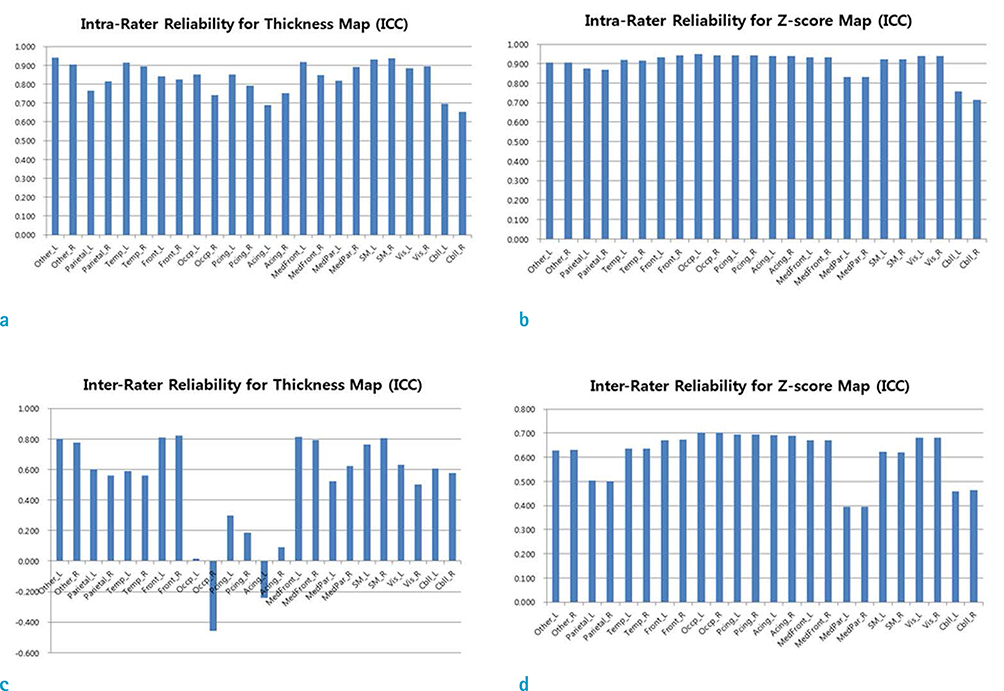
Fig. 1 Comparison of intra- and inter-rater reliability for cortical thickness and Z-scores in different brain regions.

Fig. 2 Scatter plots of cortical thickness measurements using CorThick analysis software. (a) Comparison of 2 sessions by rater 1 (R = 0.817, P < 0.01). (b) Comparison between rater 1 and rater 2 (R = 0.562, P < 0.001).

Fig. 3 Cortical thickness map (a) and Z-score map (b) using CorThick analysis software, with comparison between rater 1 and rater 2. Right lateral, left lateral, right medial, and left medial surfaces are shown (left to right).
Reference
-
1. Jack CR Jr. Alzheimer disease: new concepts on its neurobiology and the clinical role imaging will play. Radiology. 2012; 263:344–361.2. Jack CR Jr, Petersen RC, O'Brien PC, Tangalos EG. MR-based hippocampal volumetry in the diagnosis of Alzheimer's disease. Neurology. 1992; 42:183–188.3. Vermersch P, Leys D, Scheltens P, Barkhof F. Visual rating of hippocampal atrophy: correlation with volumetry. J Neurol Neurosurg Psychiatry. 1994; 57:1015.4. Ashburner J, Friston KJ. Voxel-based morphometry--the methods. Neuroimage. 2000; 11:805–821.5. Fischl B, Dale AM. Measuring the thickness of the human cerebral cortex from magnetic resonance images. Proc Natl Acad Sci U S A. 2000; 97:11050–11055.6. MacDonald D, Kabani N, Avis D, Evans AC. Automated 3-D extraction of inner and outer surfaces of cerebral cortex from MRI. Neuroimage. 2000; 12:340–356.7. Fischl B. FreeSurfer. Neuroimage. 2012; 62:774–781.8. Lin Z, Avinash G, McMillan K, Yan L, Sirohey S, Minoshima S. Quantitative measurement of MR cortical atrophy: MR brain surface intensity model (BSIM) and group and individual cortical thinning studies. In : Proc SPIE 8672, Medical Imaging 2013: Biomedical applications in molecular, structural and functional imaging, 86720E; March 29, 2013.9. Lin ZS, Avinash G, Yan L, McMillan K. Cortical thinning in cognitively normal elderly cohort of 60 to 89 year old from AIBL database and vulnerable brain areas. In : Proc SPIE 9038, Medical Imaging 2014: Biomedical applications in molecular, structural and functional imaging, 90381P; March 13, 2014.10. Lee JS, Lee DS, Kim J, et al. Development of Korean standard brain templates. J Korean Med Sci. 2005; 20:483–488.11. Tang Y, Hojatkashani C, Dinov ID, et al. The construction of a Chinese MRI brain atlas: a morphometric comparison study between Chinese and Caucasian cohorts. Neuroimage. 2010; 51:33–41.12. Chee MW, Zheng H, Goh JO, Park D, Sutton BP. Brain structure in young and old East Asians and Westerners: comparisons of structural volume and cortical thickness. J Cogn Neurosci. 2011; 23:1065–1079.13. McGraw KO WS. Forming inferences about some intraclass correlation coefficients. Psychol Methods. 1996; 1:30–46.14. Shrout PE, Fleiss JL. Intraclass correlations: uses in assessing rater reliability. Psychol Bull. 1979; 86:420–428.15. Fleiss JL. Statistical Methods for Rates and Proportions. 2nd ed. New York, NY: Wiley, John and Sons, Incorporated;1981.16. Cicchetti DV, Sparrow SA. Developing criteria for establishing interrater reliability of specific items: applications to assessment of adaptive behavior. Am J Ment Defic. 1981; 86:127–137.17. Maezawa M, Seki T, Imura S, Akiyama K, Takikawa I, Yuasa Y. Magnetic resonance signal intensity ratio of gray/white matter in children. Quantitative assessment in developing brain. Brain Dev. 1993; 15:198–204.18. Narr KL, Bilder RM, Toga AW, et al. Mapping cortical thickness and gray matter concentration in first episode schizophrenia. Cereb Cortex. 2005; 15:708–719.19. Lerch JP, Pruessner J, Zijdenbos AP, et al. Automated cortical thickness measurements from MRI can accurately separate Alzheimer's patients from normal elderly controls. Neurobiol Aging. 2008; 29:23–30.20. Du AT, Schuff N, Kramer JH, et al. Different regional patterns of cortical thinning in Alzheimer's disease and frontotemporal dementia. Brain. 2007; 130:1159–1116.21. Hartikainen P, Rasanen J, Julkunen V, et al. Cortical thickness in frontotemporal dementia, mild cognitive impairment, and Alzheimer's disease. J Alzheimers Dis. 2012; 30:857–874.22. Lehmann M, Rohrer JD, Clarkson MJ, et al. Reduced cortical thickness in the posterior cingulate gyrus is characteristic of both typical and atypical Alzheimer's disease. J Alzheimers Dis. 2010; 20:587–598.23. Ballmaier M, O'Brien JT, Burton EJ, et al. Comparing gray matter loss profiles between dementia with Lewy bodies and Alzheimer's disease using cortical pattern matching: diagnosis and gender effects. Neuroimage. 2004; 23:325–335.24. Jubault T, Gagnon JF, Karama S, et al. Patterns of cortical thickness and surface area in early Parkinson's disease. Neuroimage. 2011; 55:462–467.25. Li Y, Wang Y, Wu G, et al. Discriminant analysis of longitudinal cortical thickness changes in Alzheimer's disease using dynamic and network features. Neurobiol Aging. 2012; 33:427e415–e430.26. Thompson PM, Hayashi KM, Dutton RA, et al. Tracking Alzheimer's disease. Ann N Y Acad Sci. 2007; 1097:183–214.27. Iscan Z, Jin TB, Kendrick A, et al. Test-retest reliability of freesurfer measurements within and between sites: Effects of visual approval process. Hum Brain Mapp. 2015; 36:3472–3485.28. Clarkson MJ, Cardoso MJ, Ridgway GR, et al. A comparison of voxel and surface based cortical thickness estimation methods. Neuroimage. 2011; 57:856–865.29. Wyman BT, Harvey DJ, Crawford K, et al. Standardization of analysis sets for reporting results from ADNI MRI data. Alzheimers Dement. 2013; 9:332–337.30. Jack CR Jr, Bernstein MA, Fox NC, et al. The Alzheimer's Disease Neuroimaging Initiative (ADNI): MRI methods. J Magn Reson Imaging. 2008; 27:685–691.
- Full Text Links
-
- Actions
-
Cited
- CITED
-
- Close
- Share
-
- Similar articles
-
- Intra and Inter-Rater Measurement Reliability of Tibialis Anterior Muscle (TA) Thickness using the Ultrasonography Spring Gauge Technique
- Ultrasonographic Measurement of the Thickness of Axillary Recess Capsule in Healthy Volunteers
- The reliability of an easy measuring method for abutment convergence angle with a computer-aided design (CAD) system
- The Intra- and Inter-rater Reliability and the Learning Curve for a Simple Neurological Score for Rats
- The Validity and Reliability of the EMC Device; For the Checking ofthe Mobility of the First Ray of the Foot
