Supradiaphragmatic Liver Confirmed by a Hepatocyte-specific Contrast Agent (Gd-EOB-DTPA): A Case Report
- Affiliations
-
- 1Department of Radiology, Daejin Medical Center Bundang Jesaeng General Hospital, Seongnam-si, Gyeonggi-do, Korea. hyukjungk@naver.com
- KMID: 2175577
- DOI: http://doi.org/10.13104/imri.2015.19.1.52
Abstract
- Supradiaphragmatic liver is a rare condition. Establishing an accurate preoperative diagnosis is difficult. Operative exploration is necessary to differentiate this lesion from intrathoracic masses, such as a pleural based tumor, diaphragmatic tumor and peripheral lung tumor. However, with the aid of the hepatocyte-specific magnetic resonance imaging contrast agent, gadoxetic acid (Gd-EOB-DTPA), functional hepatocytes in the lesion can be identified in the hepatobiliary phase, potentially allowing an accurate and non-invasive diagnosis. We report a case of supradiaphragmatic liver diagnosed by Gd-EOB-DTPA-enhanced magnetic resonance imaging.
Keyword
Figure
-

Fig. 1 Supraddiaphgramatic liver in a 57-year-old woman. a-d. Dynamic liver CT images after contrast injection demonstrate a well-defined nodular mass (arrowheads) near the diaphragm. The mass shows similar dynamic enhancement to that of liver in arterial phase (a), portovenous phase (b) and delayed phase (c). Bridging vessel (arrow) from liver is visualized on coronal reformatted image of arterial phase (d).
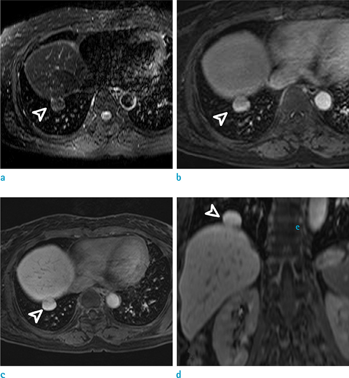
Fig. 2 Supraddiaphgramatic liver in 57-year-old woman. a. Fat saturated T2-weighted (SPIR, slice thickeness = 3 mm, TR/TE = 1964/70 msec) axial MR images demonstrate the supradiaphragmatic mass (arrowheads) with isosignal intensity comparable to liver parenchyma. b. In contrastenhanced 3D T1 weight images (Gd-EOB-DTPA, flip angle = 10°, TR/TE = 3/1.5 msec), the supradiaphragmatic mass (arrowheads) showed similar dynamic enhancement (portal phase, which is 2 minutes after contrast injection, is shown). c, d. In the hepatobiliary phase (20 minutes after contrast injection), the supradiaphragmatic mass (arrowheads) exhibits the same amount of contrast uptakes comparable to liver parenchyma. SPIR = spectral presaturation inversion recovery
Reference
-
1. Choi SU, Kim HK, Kim J. Heterotopic supradiaphragmatic liver combined with intralobar pulmonary sequestration. Ann Thorac Surg. 2008; 85:1809–1810.2. Chen YY, Huang TW, Chang H, Hsu HH, Lee SC. Intrathoracic caudate lobe of the liver: a case report and literature review. World J Gastroenterol. 2014; 20:5147–5152.3. An J, Han J, Lee KS, Choi YS. Supradiaphragmatic heterotopic liver presenting as a pleural mass: a case report. Tuberc Respir Dis. 2010; 69:191–195.4. Kurt A, Yazıcıoğlu KR, Tosun Ö, Coşkun M. Right sided diaphragmatic hernia in an adult without history of trauma: Unusual CT findings. Eur J Gen Med. 2004; 1:55–57.5. Mendoza A, Voland J, Wolf P, Benirschke K. Supradiaphragmatic liver in the lung. Arch Pathol Lab Med. 1986; 110:1085–1086.6. Agha FP. Transdiaphragmatic liver hernia in adults. Australas Radiol. 1985; 29:19–25.7. Sato K, Orihashi K, Hamanaka Y, et al. Post-traumatic diaphragmatic herniation of the liver, examined by positron emission tomography: case report. World J Emerg Surg. 2011; 6:30.8. Luoma R, Raboei E. Supradiaphragmatic accessory liver: a rare cause of respiratory distress in a neonate. J Pediatr Surg. 2003; 38:1413–1414.9. Kinnunen P, Kulmala P, Kaarteenaho-Wiik R, Vuopala K. Ectopic liver in the human pericardium. Histopathology. 1997; 30:277–279.
- Full Text Links
-
- Actions
-
Cited
- CITED
-
- Close
- Share
-
- Similar articles
-
- Hepatic Lymphoma Representing Iso-Signal Intensity on Hepatobiliary Phase, in Gd-EOB-DTPA-Enhanced MRI: Case Report
- Enhancement Pattern of Liver Parenchyma during Late Dynamic Phase Imaging: Comparison between Gd-EOB-DTPA and Gd-DTPA-BMA
- MR study of water exchange and cell membrane permeability in rat liver cells using a tissue-specific MR contrast agent
- Quantitative Evaluation of the Hepatic Parenchymal Change in Patients with Chronic Liver Disease Using Gd-EOB-DTPA-enhanced MRI: Comparison with Normal Liver
- Confident Diagnosis of Bronchobiliary Fistula Using Contrast-Enhanced Magnetic Resonance Cholangiography
