Image Findings of Sarcomatous Intrahepatic Cholangiocarcinoma Focused on Gd-EOB-DTPA Enhanced MRI: A Case Report
- Affiliations
-
- 1Department of Radiology, Daegu Fatima Hospital, Daegu, Korea. nosmokeman@naver.com
- KMID: 2175576
- DOI: http://doi.org/10.13104/imri.2015.19.1.47
Abstract
- Sarcomatous Intrahepatic cholangiocarcinoma is a rare but an aggressive variant of cholangiocarcinoma with a very poor prognosis. A 79-year-old man was admitted to our hospital because of incidentally found liver mass. Magnetic resonance imaging (MRI) revealed well-defined hypointense mass on T1WI and heterogeneous hyperintense mass on T2WI. Gd-EOB-DTPA enhanced study shows peripheral rim-like enhancement in arterial phase and progressive concentric filling of contrast in delayed phase. And mass shows significant enhancement in hepatobiliary phase. The pathologic diagnosis was intrahepatic cholangiocarcinoma with sarcomatous change.
Keyword
Figure
-
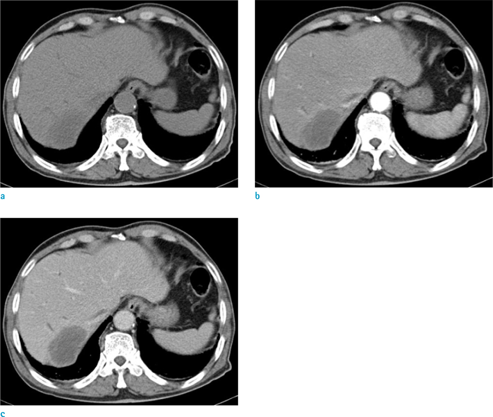
Fig. 1 A hypoattenuating mass in right posterior segment of the liver is shown on abdominal dynamic computed tomography pre (a), arterial (b) and portal phase (c) images. The mass has thin peripheral enhancement in arterial phase and concentric filling of contrast media in portal phase.

Fig. 2 Magnetic resonance (MR) imaging. An axial T1-weighted image (WI) shows a well-defined hypointense mass (a) and T2WI shows heterogeneous hyperintense mass (b) in right posterior segment of the liver. Gd-EOB-DTPA enhanced MRI obtained during the arterial phase reveals peripheral rim-like enhancement (c). The mass demonstrates progressive concentric filling of contrast in delayed phase (d) and significant enhancement in hepatobiliary phase (20 min after injection) (e). The mass shows increased SI on high-b-value diffusion-weighted image (f).
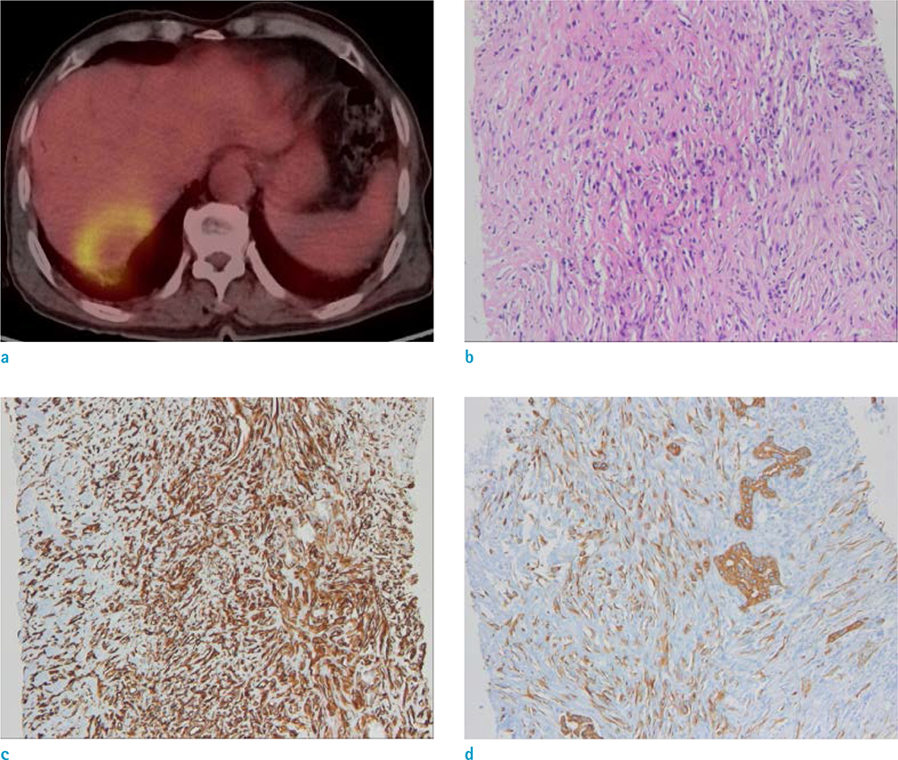
Fig. 3 Positron emission tomography (PET)-CT scan shows intense fluorodeoxyglucose (FDG) uptake in the mass, indicating possibility of malignant lesion (a). Photomicrograph (original magnification, × 100; H&E stain) shows some parts of the tumor appeared sarcomatous and were composed of pleomorphic spindle cells with numerous mitoses, whereas other parts were composed of adenocarcinoma cells (b). An immunochemical examination of the sarcomatous components showed positive staining for vimentin (c) and low molecular cytokeratin (CK7) (d).
Reference
-
1. Haratake J, Horie A. An immunohistochemical study of sarcomatoid liver carcinomas. Cancer. 1991; 68:93–97.2. Kakizoe S, Kojiro M, Nakashima T. Hepatocellular carcinoma with sarcomatous change. Clinicopathologic and immunohistochemical studies of 14 autopsy cases. Cancer. 1987; 59:310–316.3. Nakajima T, Tajima Y, Sugano I, Nagao K, Kondo Y, Wada K. Intrahepatic cholangiocarcinoma with sarcomatous change. Clinicopathologic and immunohistochemical evaluation of seven cases. Cancer. 1993; 72:1872–1877.4. Tsou YK, Wu RC, Hung CF, Lee CS. Intrahepatic sarcomatoid cholangiocarcinoma: clinical analysis of seven cases during a 15-year period. Chang Gung Med J. 2008; 31:599–605.5. Bilgin M, Toprak H, Bilgin SS, Kondakci M, Balci C. CT and MRI findings of sarcomatoid cholangiocarcinoma. Cancer Imaging. 2012; 12:447–451.6. Jeong WK, Kim YK, Song KD, Choi D, Lim HK. The MR imaging diagnosis of liver diseases using gadoxetic acid: emphasis on hepatobiliary phase. Clin Mol Hepatol. 2013; 19:360–366.7. Frydrychowicz A, Lubner MG, Brown JJ, et al. Hepatobiliary MR imaging with gadolinium-based contrast agents. J Magn Reson Imaging. 2012; 35:492–511.8. Vanderveen KA, Hussain HK. Magnetic Resonance Imaging of cholangiocarcinoma. Cancer Imaging. 2004; 4:104–115.9. Campos JT, Sirlin CB, Choi JY. Focal hepatic lesions in Gd-EOB-DTPA enhanced MRI: the atlas. Insights Imaging. 2012; 3:451–474.10. Chong YS, Kim YK, Lee MW, et al. Differentiating mass-forming intrahepatic cholangiocarcinoma from atypical hepatocellular carcinoma using gadoxetic acid-enhanced MRI. Clin Radiol. 2012; 67:766–773.
- Full Text Links
-
- Actions
-
Cited
- CITED
-
- Close
- Share
-
- Similar articles
-
- Hepatic Lymphoma Representing Iso-Signal Intensity on Hepatobiliary Phase, in Gd-EOB-DTPA-Enhanced MRI: Case Report
- Monolobar Caroli's Disease in Left Lobe of the Liver: A Case Report
- Supradiaphragmatic Liver Confirmed by a Hepatocyte-specific Contrast Agent (Gd-EOB-DTPA): A Case Report
- Quantitative Evaluation of the Hepatic Parenchymal Change in Patients with Chronic Liver Disease Using Gd-EOB-DTPA-enhanced MRI: Comparison with Normal Liver
- Clinical usefulness of T1-weighted MR cholangiography with Gd-EOB-DTPA for the evaluation of biliary complication after liver transplantation
