Is the Environment of the Endoscopy Unit a Reservoir of Pathogens?
- Affiliations
-
- 1Department of Internal Medicine, Keimyung University School of Medicine, Daegu, Korea. dandy813@hanmail.net
- 2Department of Laboratory Medicine, Keimyung University School of Medicine, Daegu, Korea.
- 3Department of Internal Medicine, Kyungpook University School of Medicine, Daegu, Korea.
- KMID: 2174408
- DOI: http://doi.org/10.5217/ir.2014.12.4.306
Abstract
- BACKGROUND/AIMS
Given the characteristic procedures involved in the endoscopy unit, the spread of pathogens is much more frequent in this unit than in other environments. However, there is a lack of data elucidating the existence of pathogens in the endoscopy unit. The aim of this study was to detect the presence of possible pathogens in the endoscopy unit.
METHODS
We performed environmental culture using samples from the endoscopy rooms of 2 tertiary hospitals. We used sterile cotton-tipped swabs moistened with sterile saline to swab the surfaces of 197 samples. Then, we cultured the swab in blood agar plate. Samples from the colonoscopy room were placed in thioglycollate broth to detect the presence of anaerobes. After 2 weeks of culture period, we counted the colony numbers.
RESULTS
The most commonly contaminated spots were the doctor's keyboard, nurse's cart, and nurse's mouse. The common organisms found were non-pathogenic bacterial microorganisms Staphylococcus, Micrococcus, and Streptococcus spp.. No definite anaerobe organism was detected in the colonoscopy room.
CONCLUSIONS
Although the organisms detected in the endoscopy unit were mainly non-pathogenic organisms, they might cause opportunistic infections in immunocompromised patients. Therefore, the environment of the endoscopy room should be managed appropriately; moreover, individual hand hygiene is important for preventing possible hospital-acquired infections.
Keyword
MeSH Terms
Figure
-
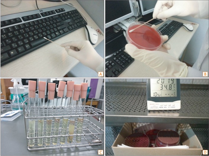
Fig. 1 The methods of environmental culture. (A) Sterile cotton-tipped swab moistened with sterile saline was used to swab the surfaces of 197 samples. (B) We cultured the swab in the blood agar plate. (C) Samples from the colonoscopy room were put in the thioglycollate broth. (D) The samples were cultured for 2 weeks.
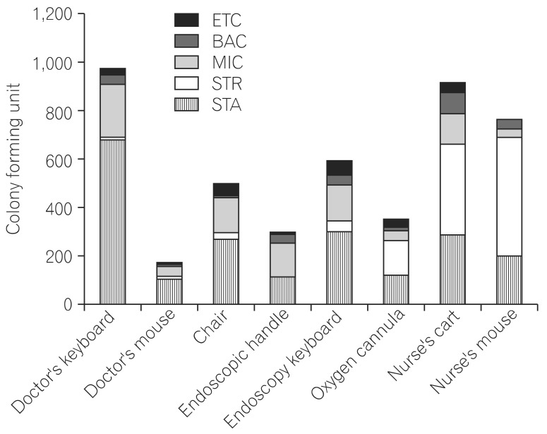
Fig. 2 Distribution of microbial species isolated by culture of samples from various surfaces. BAC, Bacillus spp.; MIC, Micrococcus spp.; STR, Streptococcus spp.; STA, staphylococcus spp.; ETC, enterococcus.

Fig. 3 Distribution of microbial species isolated by culture of samples from various surfaces. (A) Species of cultured microorganisms in the therapeutic endoscopy room. (B) Species of cultured microorganisms in the diagnostic upper endoscopy room. (C) Species of cultured microorganisms in the diagnostic colonoscopy room. BAC, Bacillus spp.; MIC, Micrococcus spp.; STR, Streptococcus spp.; STA, staphylococcus spp.; ETC, enterococcus.
Reference
-
1. Trybou J, Spaepen E, Vermeulen B, Porrez L, Annemans L. Hospital-acquired infections in Belgian acute-care hospitals: financial burden of disease and potential cost savings. Acta Clin Belg. 2013; 68:199–205. PMID: 24156220.
Article2. Pittet D, Dharan S, Touveneau S, Sauvan V, Perneger TV. Bacterial contamination of the hands of hospital staff during routine patient care. Arch Intern Med. 1999; 159:821–826. PMID: 10219927.
Article3. Hartmann B, Benson M, Junger A, et al. Computer keyboard and mouse as a reservoir of pathogens in an intensive care unit. J Clin Monit Comput. 2004; 18:7–12. PMID: 15139578.
Article4. Santos LX, Souza Dias MB, Borrasca VL, et al. Improving hand hygiene adherence in an endoscopy unit. Endoscopy. 2013; 45:421–425. PMID: 23733725.
Article5. Gamboa F, Garcia DA, Acosta A, et al. Presence and antimicrobial profile of gram-negative facultative anaerobe rods in patients with chronic periodontitis and gingivitis. Acta Odontol Latinoam. 2013; 26:24–30. PMID: 24294820.6. Dumford DM 3rd, Nerandzic MM, Eckstein BC, Donskey CJ. What is on that keyboard? Detecting hidden environmental reservoirs of Clostridium difficile during an outbreak associated with North American pulsed-field gel electrophoresis type 1 strains. Am J Infect Control. 2009; 37:15–19. PMID: 19171247.
Article7. Seo GS. Clostridium difficile Infection: What's New? Intest Res. 2013; 11:1–13.8. Bashir MH, Olson LK, Walters SA. Suppression of regrowth of normal skin flora under chlorhexidine gluconate dressings applied to chlorhexidine gluconate-prepped skin. Am J Infect Control. 2012; 40:344–348. PMID: 21737178.
Article9. Borgaonkar MR, Hookey L, Hollingworth R, et al. Indicators of safety compromise in gastrointestinal endoscopy. Can J Gastroenterol. 2012; 26:71–78. PMID: 22312605.
Article10. Appelgate D, Faust B, Dunson J. Cleaning audits lead to better environmental hygiene. OR Manager. 2013; 29:110–11.11. Matouskova I, Raida L, Holy O. The incidence of nonfermentative gram-negative bacilli in the environment of the transplant unit, department of hemato-oncology, university hospital Olomouc. Epidemiol Mikrobiol Imunol. 2012; 61:110–115. PMID: 23301626.12. Drougka E, Foka A, Marangos MN, et al. The first case of Staphylococcus aureus ST398 causing bacteremia in an immunocompromised patient in Greece. Indian J Med Microbiol. 2012; 30:232–236. PMID: 22664446.
Article13. Dwyer DM, Klein EG, Istre GR, Robinson MG, Neumann DA, McCoy GA. Salmonella newport infections transmitted by fiberoptic colonoscopy. Gastrointest Endosc. 1987; 33:84–87. PMID: 3569805.
Article14. Sabnis RB, Bhattu A, Vijaykumar M. Sterilization of endoscopic instruments. Curr Opin Urol. 2014; 24:195–202. PMID: 24451088.
Article15. Faires MC, Pearl DL, Berke O, Reid-Smith RJ, Weese JS. The identification and epidemiology of meticillin-resistant Staphylococcus aureus and Clostridium difficile in patient rooms and the ward environment. BMC Infect Dis. 2013; 13:342. PMID: 23883171.
- Full Text Links
-
- Actions
-
Cited
- CITED
-
- Close
- Share
-
- Similar articles
-
- Infection Control for Hemodialysis and Endoscopy Unit
- Contamination of the Hospital Environmental by Pathogenic Bacteria and Infection Control
- Understanding hybrid endoscopic submucosal dissection subtleties
- Traumatic Subdural Hygroma Due to Injury of an Ommaya Reservoir: A Case Report
- A Study on the Diverticular Enlargement of the Rat's Submandibular Duct (II)
