Leukemic manifestation of blastic plasmacytoid dendritic cell neoplasm: laboratory approaches in 2 cases
- Affiliations
-
- 1Department of Laboratory Medicine, Pusan National University School of Medicine, Busan, Korea.
- 2Biomedical Research Institute, Pusan National University Hospital, Busan, Korea.
- 3Department of Laboratory Medicine, University of Ulsan College of Medicine and Asan Medical Center, Seoul, Korea. hschi@amc.seoul.kr
- KMID: 2172794
- DOI: http://doi.org/10.5045/br.2014.49.3.198
Abstract
- No abstract available.
MeSH Terms
Figure
-
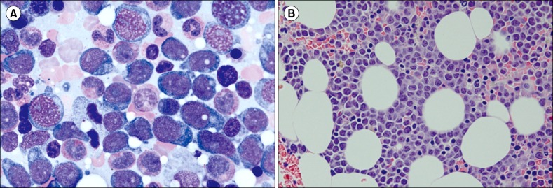
Fig. 1 Bone marrow (BM) aspiration and biopsy findings in the first case. BM aspiration showed the presence of neoplastic cells (27.8%) with oval to round nuclei, prominent nucleoli, and abundant amount of bluish cytoplasm (A, Wright staining, ×1,000). The BM clot section also showed interstitial infiltration of neoplastic cells (B, hematoxylin and eosin staining, ×400) positive for CD4, CD56, and CD123 on immunohistochemical staining.
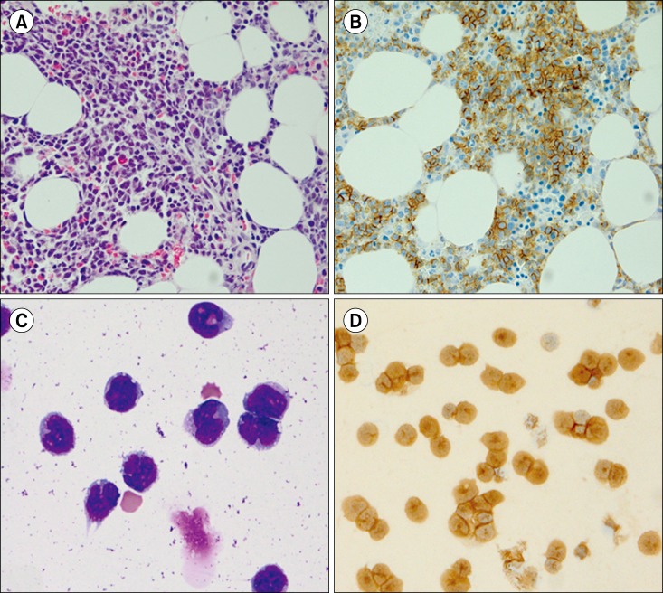
Fig. 2 Findings obtained with bone marrow biopsy and cytospin analysis of the cerebrospinal fluid in the second case. The bone marrow biopsy demonstrated an infiltration of neoplastic cells (A, hematoxylin and eosin staining, ×400) with weak positivity for CD4 and strong positivity for CD56 (B, ×400) on immunohistochemical staining. Cytospin analysis of the cerebrospinal fluid showed neoplastic cells with at frequency of 92.0% (C, Wright staining, ×400); immunocytochemical staining for both CD56 and CD123 (D, ×400) demonstrated positive neoplastic cells.
Reference
-
1. Pagano L, Valentini CG, Pulsoni A, et al. Blastic plasmacytoid dendritic cell neoplasm with leukemic presentation: an Italian multicenter study. Haematologica. 2013; 98:239–246. PMID: 23065521.
Article2. Ng AP, Lade S, Rutherford T, McCormack C, Prince HM, Westerman DA. Primary cutaneous CD4+/CD56+ hematodermic neoplasm (blastic NK-cell lymphoma): a report of five cases. Haematologica. 2006; 91:143–144. PMID: 16434387.3. Bekkenk MW, Jansen PM, Meijer CJ, Willemze R. CD56+ hematological neoplasms presenting in the skin: a retrospective analysis of 23 new cases and 130 cases from the literature. Ann Oncol. 2004; 15:1097–1108. PMID: 15205205.
Article4. Hwang K, Park CJ, Jang S, et al. Immunohistochemical analysis of CD123, CD56 and CD4 for the diagnosis of minimal bone marrow involvement by blastic plasmacytoid dendritic cell neoplasm. Histopathology. 2013; 62:764–770. PMID: 23470050.
Article5. Jacob MC, Chaperot L, Mossuz P, et al. CD4+ CD56+ lineage negative malignancies: a new entity developed from malignant early plasmacytoid dendritic cells. Haematologica. 2003; 88:941–955. PMID: 12935983.
- Full Text Links
-
- Actions
-
Cited
- CITED
-
- Close
- Share
-
- Similar articles
-
- A Woman with Blastic Plasmacytoid Dendritic Cell Neoplasm
- Blastic Plasmacytoid Dendritic Cell Neoplasm Mimicking Traumatic Hematoma: A Case Report
- A Case of Blastic Plasmacytoid Dendritic Cell Neoplasm in Child
- A Case of Blastic Plasmacytoid Dendritic Cell Neoplasm with Mutations in DNMT3A, TET2, SRSF2, and ATRX Genes
- Plasmacytoid dendritic cell neoplasms
