Chonnam Med J.
2013 Apr;49(1):48-49. 10.4068/cmj.2013.49.1.48.
Gross Hematuria Associated with Genitourinary Tuberculosis
- Affiliations
-
- 1Department of Internal Medicine, Chonnam National University Medical School, Gwangju, Korea. skimw@chonnam.ac.kr
- 2Department of Radiology, Chonnam National University Medical School, Gwangju, Korea.
- 3Department of Pathology, Chonnam National University Medical School, Gwangju, Korea.
- 4Department of Urology, Chonnam National University Medical School, Gwangju, Korea.
- KMID: 2172177
- DOI: http://doi.org/10.4068/cmj.2013.49.1.48
Abstract
- A 27-year-old man presented to the emergency department with sudden onset of massive gross hematuria and urinary retention. Contrast-enhanced computed tomography imaging showed uneven, dilated calices and a narrowing of the renal pelvis in the left kidney; in addition, a large hematoma was noted in the urinary bladder. An emergency cystoscopy was performed following detection of the hematoma and blood clots were removed. A lesional biopsy, a tuberculosis (TB) culture, and urine cytology showed positive results for Mycobacterium tuberculosis. The clinical manifestations of genitourinary tuberculosis are nonspecific and are usually detected at a chronic stage. In conclusion, we report an unusual cause of acute kidney injury associated with a subacute stage of genitourinary tuberculosis that caused mucosal erosion and bleeding in the bladder.
Keyword
MeSH Terms
Figure
-
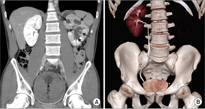
FIG. 1 (A) Contrast-enhanced excretory-phase computed tomography image showing uneven dilated calices (arrows) and narrowing of the renal pelvis (arrowhead) of the left kidney and a large hematoma in the urinary bladder. (B) Computed tomography with intravenous pyelogram image showing an invisible left pyelonephrogram compared with a normal right pyelonephrogram.
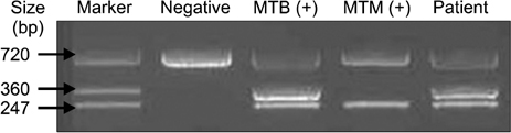
FIG. 2 Polymerase chain reaction for urine showed a positive result for Mycobacterium tuberculosis.
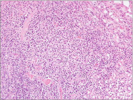
FIG. 3 Hematoxylin and eosin staining in the bladder showed caseous necrosis and inflammatory cell infiltration (×200).
Reference
-
1. Engin G, Acunaş B, Acunaş G, Tunaci M. Imaging of extrapulmonary tuberculosis. Radiographics. 2000. 20:471–488.
Article2. Burrill J, Williams CJ, Bain G, Conder G, Hine AL, Misra RR. Tuberculosis: a radiologic review. Radiographics. 2007. 27:1255–1273.
Article3. Simon HB, Weinstein AJ, Pasternak MS, Swartz MN, Kunz LJ. Genitourinary tuberculosis. Clinical features in a general hospital population. Am J Med. 1977. 63:410–420.
- Full Text Links
-
- Actions
-
Cited
- CITED
-
- Close
- Share
-
- Similar articles
-
- Clinical Observation on Gross Hematuria
- Statistical Observation of Hematuria with Urologic Diseases
- A Clinical Observation on Gross Hematuria
- Clinical Observation on In-patient of Genitourinary Tract Tuberculosis
- Influence of Gross or Microscopic Hematuria on BTA Stat Test Result in the Detection of Bladder Cancer
