Ann Dermatol.
2009 Feb;21(1):46-48. 10.5021/ad.2009.21.1.46.
A Case of Diffuse Neurofibroma of the Scalp
- Affiliations
-
- 1Department of Dermatology, College of Medicine, Chung-Ang University, Seoul, Korea. mnkim@cau.ac.kr
- 2Department of Pathology, College of Medicine, Chung-Ang University, Seoul, Korea.
- KMID: 2172065
- DOI: http://doi.org/10.5021/ad.2009.21.1.46
Abstract
- A neurofibroma is a benign tumor of the peripheral nerve sheath characterized by proliferation of Schwann cells, perineural cells, and endoneurial fibroblasts. Different types of neurofibromas can be identified, including localized, plexiform, and diffuse types. Neurofibromas can involve any site on the body skin. The diffuse variant is rare and occurs primarily in children and young adults. It involves the skin and subcutaneous tissue in a plaque-like fashion on the head and neck regions. We present a case of a 10-year-old boy who had a diffuse neurofibroma on the scalp.
Keyword
MeSH Terms
Figure
-
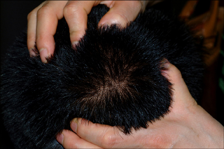
Fig. 1 A 7×8 cm swollen mass on the patient's scalp.

Fig. 2 Radiologic findings. (A) The MRI scan. The axial T1-weighted spin-echo image (TR/TE, 483/12) shows thickening with intermediate signal intensity in the right high frontoparietal scalp. (B) The MRI scan. The post-contrast sagittal T1-weighted spin-echo image shows marked enhancement of the lesion in the right high frontoparietal scalp.
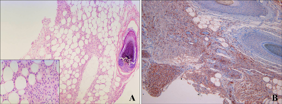
Fig. 3 (A) Histopathologic examination showed fusiform cells with elongated nuclei in a myxoid matrix with wire-like collagen fibers (H&E, ×100, inset, ×400). (B) Immunoperoxidase staining for S-100 protein was positive (S-100, ×100).
Reference
-
1. Peh WC, Shek TW, Yip DK. Magnetic resonance imaging of subcutaneous diffuse neurofibroma. Br J Radiol. 1997. 70:1180–1183.
Article2. Yang JS, Park HJ, Lee HJ, Baek SC, Byun DG. A case of trichotillomania associated with diffuse neurofibroma. Korean J Dermatol. 2001. 39:1152–1156.3. Cha SH, Cho SH, Lee JD. A case of diffuse neurofibroma. Korean J Dermatol. 2008. 46:355–358.4. Daoud MS, Pittelkow MR. Freedberg IM, Eisen AZ, Wolff K, Austen KF, Goldsmith LA, Katz SI, editors. Lichen planus. Fitzpatrick's dermatology in general medicine. 2003. 6th ed. New York: McGraw-Hill;463–477.5. van Zuuren EJ, Posma AN. Diffuse neurofibroma on the lower back. J Am Acad Dermatol. 2003. 48:938–940.
Article6. Ergun SS, Emel E, Karabekir S, Buyukbabani N. Extracranial diffuse neurofibroma with intracranial extension. Plast Reconstr Surg. 2000. 105:801–803.
Article7. Megahed M. Histopathological variants of neurofibroma. A study of 114 lesions. Am J Dermatopathol. 1994. 16:486–495.8. Kransdorf MJ, Berquist TH. Berquist TH, editor. Neurofibroma. MRI of the musculoskeletal system. 1996. 3rd ed. Philadelphia: Lippincott-Raven;789–792.9. Ito H, Akagi O, Nomura N, Tahara E. Giant pigmented tumour of the scalp--a diffuse neurofibroma or a congenital naevus showing neurofibromatous changes? Immunohistochemical and electron microscopic studies. Histopathology. 1988. 13:181–189.
Article10. Beggs I, Gilmour HM, Davie RM. Diffuse neurofibroma of the ankle. Clin Radiol. 1998. 53:755–759.
Article
