Ann Dermatol.
2011 Aug;23(3):415-416. 10.5021/ad.2011.23.3.415.
Isolated Epidermolytic Acanthoma in a Renal Transplant Recipient
- Affiliations
-
- 1Department of Dermatology, Asan Medical Center, University of Ulsan College of Medicine, Seoul, Korea. miumiu@amc.seoul.kr
- KMID: 2171944
- DOI: http://doi.org/10.5021/ad.2011.23.3.415
Abstract
- No abstract available.
MeSH Terms
Figure
-
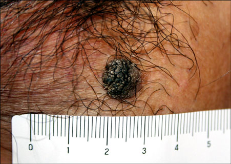
Fig. 1 A solitary, brown papule, measured 1 cm in diameter, with a verrucous surface on the scrotum.
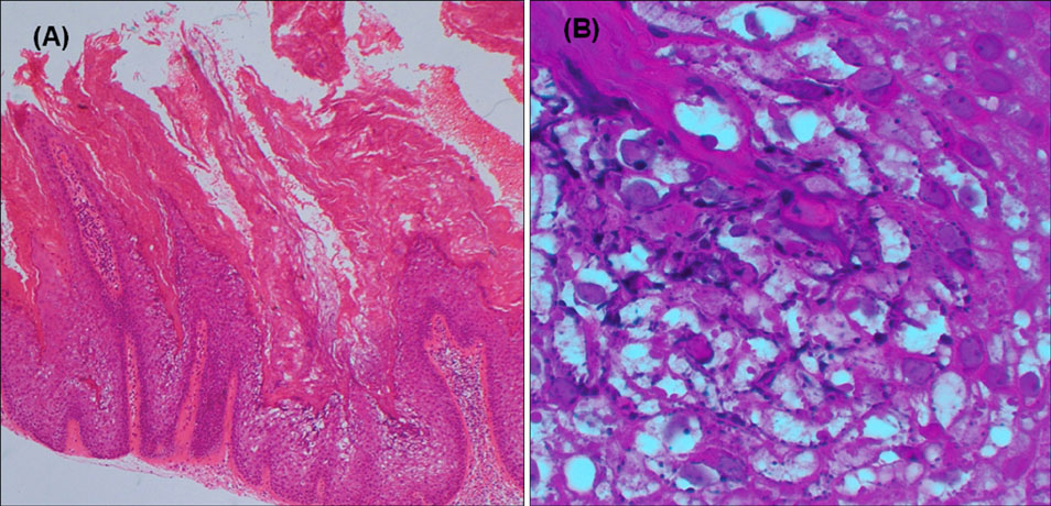
Fig. 2 Histologic examination of the scrotal lesion showing epidermolytic hyperkeratosis: vacuolar degeneration, increased keratohyaline-like bodies, hypergranulosis and compact hyperkeratosis (A: H&E, ×40, B: H&E, ×400).
Reference
-
1. Shapiro L, Baraf CS. Isolated epidermolytic acanthoma. A solitary tumor showing granular degeneration. Arch Dermatol. 1970. 101:220–223.
Article2. Ackerman AB. Histopathologic concept of epidermolytic hyperkeratosis. Arch Dermatol. 1970. 102:253–259.
Article3. Cohen PR, Ulmer R, Theriault A, Leigh IM, Duvic M. Epidermolytic acanthomas: clinical characteristics and immunohistochemical features. Am J Dermatopathol. 1997. 19:232–241.
Article4. Leonardi C, Zhu W, Kinsey W, Penneys NS. Epidermolytic acanthoma does not contain human papillomavirus DNA. J Cutan Pathol. 1991. 18:103–105.
Article5. Chun SI, Lee JS, Kim NS, Park KD. Disseminated epidermolytic acanthoma with disseminated superficial porokeratosis and verruca vulgaris in an immunosuppressed patient. J Dermatol. 1995. 22:690–692.
Article6. Nakagawa T, Nishimoto M, Takaiwa T. Disseminated epidermolytic acanthoma revealed by PUVA. Dermatologica. 1986. 173:150–153.
Article
