Ann Dermatol.
2011 Aug;23(3):379-381. 10.5021/ad.2011.23.3.379.
A Case of Myxoid Dermatofibrosarcoma Protuberans
- Affiliations
-
- 1Department of Dermatology, School of Medicine, Ewha Womans University, Seoul, Korea. hychoi@ewha.ac.kr
- KMID: 2171934
- DOI: http://doi.org/10.5021/ad.2011.23.3.379
Abstract
- Dermatofibrosarcoma protuberans (DFSP) is a slowly growing dermal spindle cell tumor and its myxoid variant, a rare type of DFSP, is characterized by extensive myxoid degeneration. We present the case of a 69-year-old woman with a multinodular reddish plaque on her trunk. Histopathologically, the tumor was located in the dermis and consisted of uniform spindle-shaped cells, showing strongly positive reaction for CD34, and negative for both S-100 and desmin. In addition to the typical storiform pattern, prominent myxoid stromal changes were demonstrated. Herein, we report an interesting case of myxoid DFSP, rarely reported in the dermatology literature.
Figure
-

Fig. 1 Multinodular reddish plaques, with partial gelatinous appearance on the abdomen.

Fig. 2 The diffusely infiltrating tumor was located in the entire dermis (H&E, ×40).
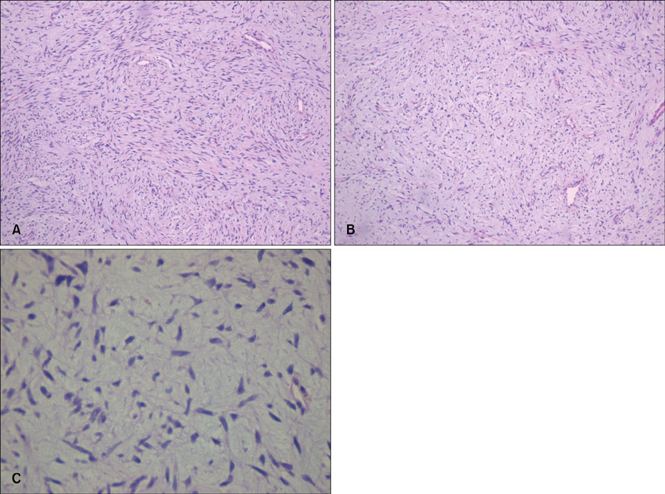
Fig. 3 (A) The tumor showed the characteristic storiform pattern in some parts of the lesion (H&E, ×100). (B) In addition to the typical storiform pattern, prominent myxoid stromal changes were demonstrated (H&E, ×100). (C) Bland spindle cells with oval nuclei were randomly embedded in the loose myxoid stroma (H&E, ×400).
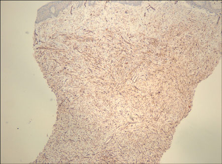
Fig. 4 (A) The tumor cells were strongly positive for CD34 (CD34, ×40).
Reference
-
1. McKee PH, Calonje JE, Granter S, Calonje E, Granter SR. Pathology of the skin: with clinical correlations. 2005. Vol 2. Philadelphia, Pa: Elsevier Mosby.2. Graadt van Roggen JF, Hogendoorn PC, Fletcher CD. Myxoid tumours of soft tissue. Histopathology. 1999. 35:291–312.
Article3. Reimann JD, Fletcher CD. Myxoid dermatofibrosarcoma protuberans: a rare variant analyzed in a series of 23 cases. Am J Surg Pathol. 2007. 31:1371–1377.
Article4. Haycox CL, Odland PB, Olbricht SM, Piepkorn M. Immunohistochemical characterization of dermatofibrosarcoma protuberans with practical applications for diagnosis and treatment. J Am Acad Dermatol. 1997. 37:438–444.
Article5. Mentzel T, Schärer L, Kazakov DV, Michal M. Myxoid dermatofibrosarcoma protuberans: clinicopathologic, immunohistochemical, and molecular analysis of eight cases. Am J Dermatopathol. 2007. 29:443–448.
Article6. Mertens F, Fletcher CD, Antonescu CR, Coindre JM, Colecchia M, Domanski HA, et al. Clinicopathologic and molecular genetic characterization of low-grade fibromyxoid sarcoma, and cloning of a novel FUS/CREB3L1 fusion gene. Lab Invest. 2005. 85:408–415.
Article
- Full Text Links
-
- Actions
-
Cited
- CITED
-
- Close
- Share
-
- Similar articles
-
- A Case of Dermatofibrosarcoma Protuberans with Myxoid Area
- Comments to “Pigmented Dermatofibrosarcoma Protuberans Presenting as a Faint Blue Macule in a Middle-aged Korean Womanâ€
- A Case of Dermatofibrosarcoma Protuberans as a Subcutaneous Nodule without Surface Change
- A Case of Dermatofibrosarcoma Protuberans Treated with Slow Mohs Micrographic Surgery
- Dermatofibrosarcoma Protuberans Arising froma Burn Scar
