The Immunohistochemical Patterns of Calcification-related Molecules in the Epidermis and Dermis of the Zebrafish (Danio rerio)
- Affiliations
-
- 1Department of Dermatology, School of Medicine, Ewha Womans University, Seoul, Korea. hychoi@ewha.ac.kr
- KMID: 2171920
- DOI: http://doi.org/10.5021/ad.2011.23.3.299
Abstract
- BACKGROUND
The scales of bony fish represent a significant reservoir of calcium and calcification of the elasmoid scale is known to be associated with deposition of mineral crystals from the epidermis to dermis. However, little is known about the exact mechanisms of calcium deposition, mobilization and regeneration occurring in the zebrafish skin.
OBJECTIVE
The purpose of this study was to investigate the expression of calcification-related molecular mediators in both the epidermis and dermis of the zebrafish (Danio rerio), using immunohistochemical study.
METHODS
We examined the skin of zebrafish in four populations of different ages (i.e. 20 days post-fertilization (dpf), 35 dpf, 50 dpf, and the adult zebrafish), using several immunohistochemical markers, including bone morphogenetic protein 4 (BMP-4), beta-catenin, osteocalcin, osteopontin and osteonectin.
RESULTS
BMP-4, osteopontin and osteonectin were moderately expressed in the epidermis of zebrafish after 35 dpf. Also, some of the cells in the upper dermis showed strong positivity for BMP-4, osteocalcin, osteopontin and osteonetin.
CONCLUSION
Our results suggest that BMP-4, osteocalcin, osteopontin and osteonectin may play a role in the process of calcification of the elasmoid scale.
Keyword
MeSH Terms
Figure
-

Fig. 1 Semi-thin sections from the zebrafish skin. (A) 7 weeks. The 2 layer-thick epidermis (E) covers the dermis (D), which is composed of a relatively homogenous layer. (B) 9 months. Under the epidermis, well-developed collagen and elastic fibers (arrow) are seen in dermis. There is a thin, homogenous layer just below the epidermis, which is called the elasmoid scale (arrowhead) in zebrafish (Toluidine Blue stain, ×400).
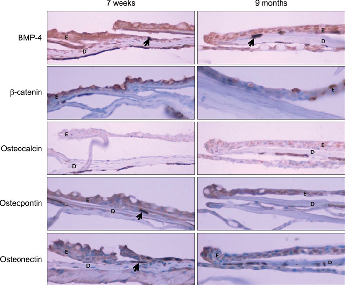
Fig. 2 Immunohistochemical stain for BMP-4, β-catenin, osteocalcin, osteopontin and osteonectin in 7 week-old and 9 month-old zebrafish skin. After 7 weeks, the epidermis (E) showed distinct positivity for BMP-4, osteopontin and osteonectin and some of the cells (arrows) in the dermis (D) also strongly expressed BMP-4, osteopontin and osteonectin (×400).
Reference
-
1. Detrich HW. Detrich HW, Westerfield M, Zon LI, editors. Overview of the zebrafish system. The zebrafish: biology in methods in cell biology. 1999. San Diego: Academic Press;5.2. Tingaud-Sequeira A, Forgue J, André M, Babin PJ. Epidermal transient down-regulation of retinol-binding protein 4 and mirror expression of apolipoprotein Eb and estrogen receptor 2a during zebrafish fin and scale development. Dev Dyn. 2006. 235:3071–3079.
Article3. Sire JY, Akimenko MA. Scale development in fish: a review, with description of sonic hedgehog (shh) expression in the zebrafish (Danio rerio). Int J Dev Biol. 2004. 48:233–247.
Article4. Sire JY, Allizard F, Babiar O, Bourguignon J, Quilhac A. Scale development in zebrafish (Danio rerio). J Anat. 1997. 190:545–561.
Article5. Sire JY, Quilhac A, Bourguignon J, Allizard F. Evidence for participation of the epidermis in the deposition of superficial layer of scales in zebrafish (Danio rerio). J Morphol. 1997. 231:161–174.
Article6. Waterman RE. Fine structure of scale development in the teleost, Brachydanio rerio. Anat Rec. 1970. 168:361–379.7. Kim SY, Choi HY, Myung KB, Choi YW. The expression of molecular mediators in the idiopathic cutaneous calcification and ossification. J Cutan Pathol. 2008. 35:826–831.
Article8. Urganus AL, Zhao YD, Pachman LM. Juvenile dermatomyositis calcification selectively displayed markers of bone formation. Arthritis Rheum. 2009. 61:501–508.
Article9. Marhaug G, Shah V, Shroff R, Varsani H, Wedderburn LR, Pilkington CA, et al. Age-dependent inhibition of ectopic calcification: a possible role for fetuin-A and osteopontin in patients with juvenile dermatomyositis with calcinosis. Rheumatology (Oxford). 2008. 47:1031–1037.
Article10. Davies CA, Jeziorska M, Freemont AJ, Herrick AL. Expression of osteonectin and matrix Gla protein in scleroderma patients with and without calcinosis. Rheumatology (Oxford). 2006. 45:1349–1355.
Article11. Reddi AH. Role of morphogenetic proteins in skeletal tissue engineering and regeneration. Nat Biotechnol. 1998. 16:247–252.
Article12. Mikhaylova L, Malmquist J, Nurminskaya M. Regulation of in vitro vascular calcification by BMP4, VEGF and Wnt3a. Calcif Tissue Int. 2007. 81:372–381.
Article13. Li G, Corsi-Payne K, Zheng B, Usas A, Peng H, Huard J. The dose of growth factors influences the synergistic effect of vascular endothelial growth factor on bone morphogenetic protein 4-induced ectopic bone formation. Tissue Eng Part A. 2009. 15:2123–2133.
Article14. Johnson ML, Rajamannan N. Diseases of Wnt signaling. Rev Endocr Metab Disord. 2006. 7:41–49.
Article15. Gadeau AP, Chaulet H, Daret D, Kockx M, Daniel-Lamazière JM, Desgranges C. Time course of osteopontin, osteocalcin, and osteonectin accumulation and calcification after acute vessel wall injury. J Histochem Cytochem. 2001. 49:79–86.
Article
- Full Text Links
-
- Actions
-
Cited
- CITED
-
- Close
- Share
-
- Similar articles
-
- Cloning of Danio rerio toll-like receptor 1
- Electron-microscopic Findings of Elastic Fibers in Zebrafish Skin
- Acute toxicity assessment of camphor in biopesticides by using Daphnia magna and Danio rerio
- Chronic Reserpine Administration for Depression Modeling in Zebrafish
- Ultrastructure and Development of Heart Wall of Zebrafish (Danio rerio)
