Ann Dermatol.
2013 May;25(2):251-252. 10.5021/ad.2013.25.2.251.
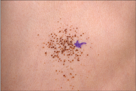
Multiple Agminated Acquired Melanocytic Nevi
- Affiliations
-
- 1Department of Dermatology, Ajou University School of Medicine, Suwon, Korea. maychan@ajou.ac.kr
- KMID: 2171802
- DOI: http://doi.org/10.5021/ad.2013.25.2.251
Abstract
- No abstract available.
MeSH Terms
Figure
Reference
-
1. Happle R. Segmental lesions are not always agminated. Arch Dermatol. 2002. 138:838.
Article2. Betti R, Inselvini E, Palvarini M, Crosti C. Agminated intradermal Spitz nevi arising on an unusual speckled lentiginous nevus with localized lentiginosis: a continuum? Am J Dermatopathol. 1997. 19:524–527.
Article3. Schaffer JV, Orlow SJ, Lazova R, Bolognia JL. Speckled lentiginous nevus: within the spectrum of congenital melanocytic nevi. Arch Dermatol. 2001. 137:172–178.4. Stewart DM, Altman J, Mehregan AH. Speckled lentiginous nevus. Arch Dermatol. 1978. 114:895–896.
Article5. Corradin MT, Alaibac M, Fortina AB. A case of malignant melanoma arising from an acquired agminated melanocytic naevus. Acta Derm Venereol. 2007. 87:432–433.
Article
- Full Text Links
- Actions
-
Cited
- CITED
-
- Close
- Share
- Similar articles
-
- Acquired Generalized Blue Nevi
- Immunohistochemical Expression of bcl-2 and PCNA in Acquired Melanocytic Nevi
- A Case of Multiple Agminated Spitz Nevi Showing Desmoplastic Changes
- A Case of Congenital Melanocytic Nevi with Unusual Cutaneous Manifestation
- Agminated Acquired Melanocytic Nevi of the Common and Dysplastic Type


