Onychomycosis Caused by Chaetomium globosum
- Affiliations
-
- 1Department of Dermatology, College of Medicine, Dongguk University, Gyeongju, Korea. smg@dongguk.ac.kr
- 2Department of Laboratory Medicine, College of Medicine, Dongguk University, Gyeongju, Korea.
- 3Department of Microbiology, College of Medicine, Dongguk University, Gyeongju, Korea.
- 4Department of Dermatology, College of Medicine, Yeungnam University, Daegu, Korea.
- KMID: 2171796
- DOI: http://doi.org/10.5021/ad.2013.25.2.232
Abstract
- Onychomycosis is usually caused by dermatophytes, but some nondermatophytic molds and yeasts are also associated with invasion of nails. The genus Chaetomium is a dematiaceous nondermatophytic mold found in soil and plant debris as a saprophytic fungus. We report the first Korean case of onychomycosis caused by Chaetomium globosum in a 35-year-old male. The patient showed brownish-yellow discoloration and subungual hyperkeratosis on the right toenails (1st and 5th) and left toenails (1st and 4th). Direct microscopic examination of scraping on the potassium hydroxide preparation revealed septate hyphae and repeated cultures on Sabouraud's dextrose agar (SDA) without cycloheximide slants showed the same fast-growing colonies, which were initially velvety white then turned to dark gray to brown. However, there was no growth of colony on SDA with cycloheximide slants. Brown-colored septated hyphae, perithecia and ascospores were shown in the slide culture. The DNA sequence of internal transcribed spacer region of the clinical sample was a 100% match to that of C. globosum strain ATCC 6205 (GenBank accession number EF524036.1). We confirmed C. globosum by KOH mount, colony, and light microscopic morphology and DNA sequence analysis. The patient was treated with 250 mg oral terbinafine daily and topical amorolfine 5% nail lacquer for 3 months.
Keyword
MeSH Terms
-
Agar
Arthrodermataceae
Base Sequence
Chaetomium
Cycloheximide
Fungi
Glucose
Humans
Hydroxides
Hyphae
Lacquer
Light
Male
Morpholines
Nails
Naphthalenes
Onychomycosis
Plants
Potassium
Potassium Compounds
Sequence Analysis, DNA
Soil
Sprains and Strains
Yeasts
Agar
Cycloheximide
Glucose
Hydroxides
Morpholines
Naphthalenes
Potassium
Potassium Compounds
Soil
Figure
-
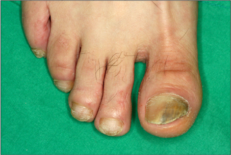
Fig. 1 Brownish-yellow discoloration with hyperkeratosis on the right toenails.

Fig. 2 Close up view of the right first toenail.
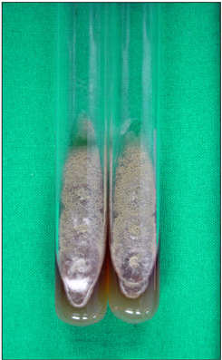
Fig. 3 Multiple, dark grey to brown colonies with aerial mycelium on Sabouraud's dextrose agar slants after incubation at 25℃ for 1 week.

Fig. 4 A rapid growing, dark grey to brown colony with aerial mycelium on Sabouraud's dextrose agar plate after incubation at 25℃ for 1 week.
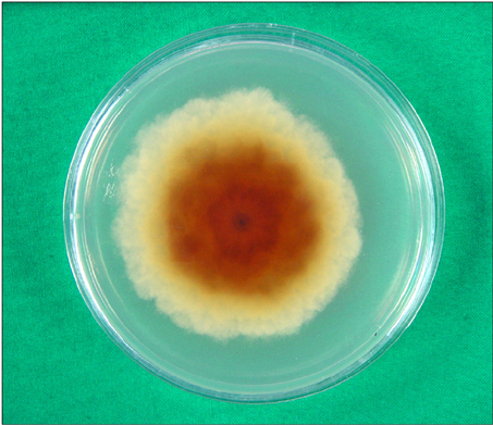
Fig. 5 Reverse surface of Sabouraud's dextrose agar plate after incubation at 25℃ for 1 week.
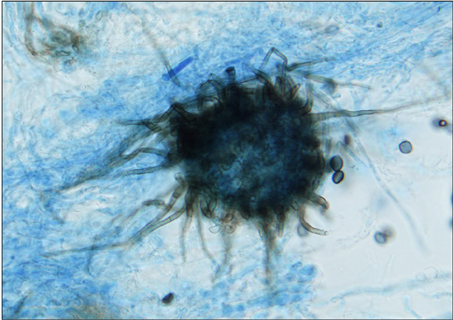
Fig. 6 Large, dark brown to black, flask shaped perithecia with hair-like filamentous appendages (Lactophenol cotton blue stain, ×400).
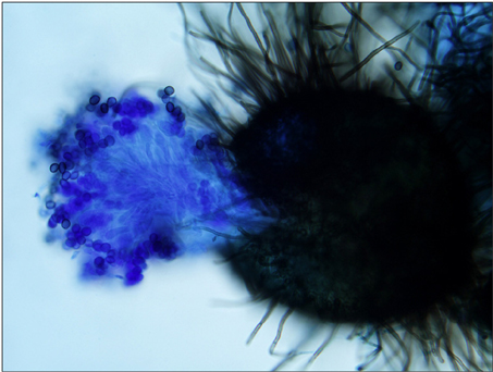
Fig. 7 Close-up view of perithecium which demonstrates ostioles and contains asci and single-celled ascospores (Lactophenol cotton blue stain, ×200).

Fig. 8 Brown-colored septated hyphae and lemon-shaped ascospores (Lactophenol cotton blue stain, ×400).
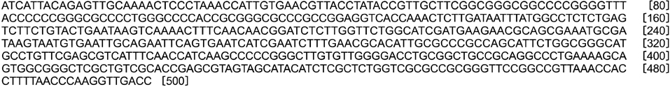
Fig. 9 Alignment of internal transcribed spacer (ITS) sequences of the sample from the patient and Chaetomium globosum strain ATCC 6205 (GenBank accession number EF524036.1). The sequences of ITS for the clinical sample was a 100% match to that of C. globosum strain ATCC 6205 (GenBank accession number EF524036.1).
Reference
-
1. Verma S, Heffernan MP. Wolff K, Goldsmith LA, Katz SI, Gilchrest BA, Paller AS, Leffell DJ, editors. Superficial fungal infection: dermatophytosis, onychomycosis, tinea nigra, piedra. Fitzpatrick's dermatology in general medicine. 2008. 7th ed. New York: McGraw-Hill;1807–1821.2. Gianni C, Cerri A, Crosti C. Non-dermatophytic onychomycosis. An understimated entity? A study of 51 cases. Mycoses. 2000. 43:29–33.
Article3. Gupta AK, Ryder JE, Baran R, Summerbell RC. Non-dermatophyte onychomycosis. Dermatol Clin. 2003. 21:257–268.
Article4. Falcón CS, Falcón Ma del MS, Ceballos JD, Florencio VD, Erchiga VC, Ortega SS. Onychomycosis by Chaetomium spp. Mycoses. 2009. 52:77–79.5. Rippon JW. Medical mycology: the pathogenic fungi and the pathogenic actinomycetes. 1988. 3rd ed. Philadelphia: WB Saunders;213.6. Naidu J, Singh SM, Pouranik M. Onychomycosis caused by Chaetomium globosum Kunze. Mycopathologia. 1991. 113:31–34.
Article7. Stiller MJ, Rosenthal S, Summerbell RC, Pollack J, Chan A. Onychomycosis of the toenails caused by Chaetomium globosum. J Am Acad Dermatol. 1992. 26:775–776.
Article8. Hattori N, Adachi M, Kaneko T, Shimozuma M, Ichinohe M, Iozumi K. Case report. Onychomycosis due to Chaetomium globosum successfully treated with itraconazole. Mycoses. 2000. 43:89–92.
Article9. Aspiroz C, Gené J, Rezusta A, Charlez L, Summerbell RC. First Spanish case of onychomycosis caused by Chaetomium globosum. Med Mycol. 2007. 45:279–282.
Article10. Latha R, Sasikala R, Muruganandam N, Shiva Prakash MR. Onychomycosis due to ascomycete Chaetomium globosum: a case report. Indian J Pathol Microbiol. 2010. 53:566–567.
Article11. Gianni C, Romano C. Clinical and histological aspects of toenail onychomycosis caused by Aspergillus spp.: 34 cases treated with weekly intermittent terbinafine. Dermatology. 2004. 209:104–110.
Article12. Tosti A, Piraccini BM, Lorenzi S. Onychomycosis caused by nondermatophytic molds: clinical features and response to treatment of 59 cases. J Am Acad Dermatol. 2000. 42:217–224.
Article13. Summerbell RC, Kane J, Krajden S. Onychomycosis, tinea pedis and tinea manuum caused by non-dermatophytic filamentous fungi. Mycoses. 1989. 32:609–619.
Article14. Arrese JE, Piérard-Franchimont C, Piérard GE. Unusual mould infection of the human stratum corneum. J Med Vet Mycol. 1997. 35:225–227.
Article15. English MP. Nails and fungi. Br J Dermatol. 1976. 94:697–701.
Article16. Yu J, Yang S, Zhao Y, Li R. A case of subcutaneous phaeoPhyphomycosis caused by Chaetomium globosum and the sequences analysis of C. globosum. Med Mycol. 2006. 44:541–545.
Article17. Tullio V, Banche G, Allizond V, Roana J, Mandras N, Scalas D, et al. Non-dermatophyte moulds as skin and nail foot mycosis agents: Phoma herbarum, Chaetomium globosum and Microascus cinereus. Fungal Biol. 2010. 114:345–349.
Article18. Nolting S, Brautigam M, Weidinger G. Terbinafine in onychomycosis with involvement by non-dermatophytic fungi. Br J Dermatol. 1994. 130:Suppl 43. 16–21.
Article19. Guarro J, Soler L, Rinaldi MG. Pathogenicity and antifungal susceptibility of Chaetomium species. Eur J Clin Microbiol Infect Dis. 1995. 14:613–618.
- Full Text Links
-
- Actions
-
Cited
- CITED
-
- Close
- Share
-
- Similar articles
-
- Descriptive Reports on Some Soil-Inhabiting Fungi in Korea
- Onychomycosis Due to Nondermatophytic Molds
- Cellulose Utilization and Protein Productivity of Some Cellulolytic Fungal Co-cultures
- Biological Control of Phytophthora palmivora Causing Root Rot of Pomelo Using Chaetomium spp.
- Phyllosphere and Phylloplane Fungi of Banana Cultivated in Upper Egypt and their Cellulolytic Ability
