Ann Dermatol.
2014 Apr;26(2):278-279. 10.5021/ad.2014.26.2.278.
Infantile Perianal Pyramidal Protrusion
- Affiliations
-
- 1Department of Dermatology, Ajou University School of Medicine, Suwon, Korea. maychan@ajou.ac.kr
- KMID: 2171676
- DOI: http://doi.org/10.5021/ad.2014.26.2.278
Abstract
- No abstract available.
Figure
-
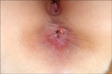
Fig. 1 Solitary skin-colored soft papule is noted on the anterior part of the anus.
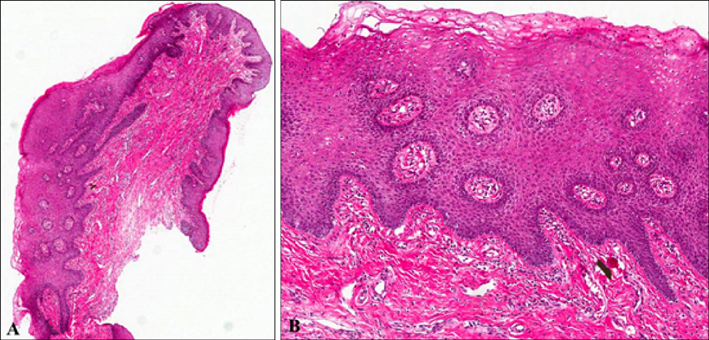
Fig. 2 (A) A papular lesion containing fibrovascular core with inflammatory cells infiltration is noted at a lower magnification. (B) Epidermal acanthosis and dilated vessels with fibrous tissue infiltrated by lymphocytes are mainly observed at higher magnification. A: H&E, ×10; B: H&E, ×100.
Reference
-
1. Zavras N, Christianakis E, Tsamoudaki S, Velaoras K. Infantile perianal pyramidal protrusion: a report of 8 new cases and a review of the literature. Case Rep Dermatol. 2012; 4:202–206.
Article2. Kim BJ, Woo SM, Li K, Lee DH, Cho S. Infantile perianal pyramidal protrusion treated by topical steroid application. J Eur Acad Dermatol Venereol. 2007; 21:263–264.3. Strand A, Andersson S, Zehbe I, Wilander E. HPV prevalence in anal warts tested with the MY09/MY11 SHARP Signal system. Acta Derm Venereol. 1999; 79:226–229.
Article4. Kaidar-Person O, Person B, Wexner SD. Hemorrhoidal disease: A comprehensive review. J Am Coll Surg. 2007; 204:102–117.
Article5. Laurence AE, Murray AJ. Histopathology of prolapsed and thrombosed hemorrhoids. Dis Colon Rectum. 1962; 5:56–61.
Article
