Piebaldism with Neurofibromatosis Type I: A Familial Case
- Affiliations
-
- 1Department of Dermatology, Yonsei University Wonju College of Medicine, Wonju, Korea. ahnsk@yonsei.ac.kr
- 2Department of Dermatology, Atopy and Asthma Center, Seoul Medical Center, Seoul, Korea.
- KMID: 2171670
- DOI: http://doi.org/10.5021/ad.2014.26.2.264
Abstract
- No abstract available.
MeSH Terms
Figure
-
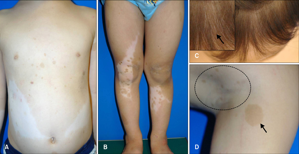
Fig. 1 The patient presented with depigmented patches and café-au-lait macules on the abdomen and extremities. (A, B) Hyperpigmented macules were present within the leukodermic patches. (C) He also had the characteristic white forelock. The arrow indicates the white forelock. (D) Multiple café-au-lait macules and freckling were noted in the axillary areas. The arrow indicates the café-au-lait macules, while the dotted lines indicate the freckling.

Fig. 2 Skin biopsies were obtained from a café-au-lait macule and a depigmented lesion. Basal hyperpigmentation was present in the café-au-lait macule (A) but not in the depigmented area (B). Fontana-Masson stain revealed increased melanin pigments in the basal layer of the specimen from the café-au-lait macule (C) but not the depigmented area specimen (D). (E) S-100-positive dendritic epidermal cells were detected in the pigmented macule. The S-100-positive cells are indicated with black arrows. (F) In the leukodermic area, some melanin pigments were deposited in the epidermis. (G) The pedigree shows family members affected with piebaldism or neurofibromatosis type 1 (NF-1). Members with only depigmented patches are marked with black boxes, while those who had both piebaldism and NF-1 are marked with deviant crease lines. The arrow indicates the patient (A, B: H&E, ×200; C, D: Fontana-Masson stain, ×200; E, F: immunohistochemical stain for S-100, ×400).
Cited by 1 articles
-
Piebaldism Associated with Café-au-lait Macules and Intertriginous Freckling: A Case Report and Review of the Literature
Sevgi Akarsu, Turna İlknur, Ceylan Avcı, Emel Fetil
Ann Dermatol. 2019;31(5):567-570. doi: 10.5021/ad.2019.31.5.567.
Reference
-
1. Richards KA, Fukai K, Oiso N, Paller AS. A novel KIT mutation results in piebaldism with progressive depigmentation. J Am Acad Dermatol. 2001; 44:288–292.
Article2. Thomas I, Kihiczak GG, Fox MD, Janniger CK, Schwartz RA. Piebaldism: an update. Int J Dermatol. 2004; 43:716–719.
Article3. Neurofibromatosis. Conference statement. National Institutes of Health Consensus Development Conference. Arch Neurol. 1988; 45:575–578.4. Tay YK. Neurofibromatosis 1 and piebaldism: a case report. Dermatology. 1998; 197:401–402.
Article5. Chang T, McGrae JD Jr, Hashimoto K. Ultrastructural study of two patients with both piebaldism and neurofibromatosis 1. Pediatr Dermatol. 1993; 10:224–234.
Article
- Full Text Links
-
- Actions
-
Cited
- CITED
-
- Close
- Share
-
- Similar articles
-
- Piebaldism Associated with Café-au-lait Macules and Intertriginous Freckling: A Case Report and Review of the Literature
- Piebaldism: a case report
- A Case of Piebaldism Associated with Strabismus and Torticollis
- A Case of Orbital Neurilemoma Associated with Neurofibroma tosis
- Four Generations of Piebaldism
