Ann Dermatol.
2015 Oct;27(5):629-630. 10.5021/ad.2015.27.5.629.
A Case of Focal Eosinophilic Myositis Associated with Hypereosinophilic Syndrome: A Rare Case Report
- Affiliations
-
- 1Department of Dermatology, Asan Medical Center, University of Ulsan College of Medicine, Seoul, Korea. miumiu@amc.seoul.kr
- KMID: 2171472
- DOI: http://doi.org/10.5021/ad.2015.27.5.629
Abstract
- No abstract available.
Figure
-
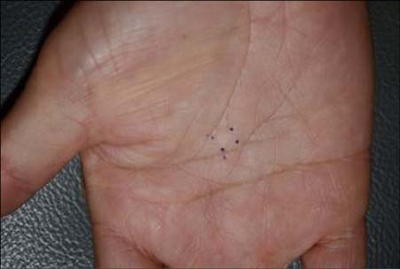
Fig. 1 A solitary well demarcated erythematous to skin colored ovoid subcutaneous nodule on the right palm.
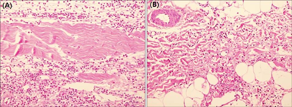
Fig. 2 (A) The marked infiltrates composed of numerous eosinophis with a slight admixture of lymphocytes in the perimysium (H&E, ×200). (B) The moderate infiltrates of eosinophils and lymphocytes in the subcutaneous fat layer (H&E, ×200).
Reference
-
1. Plötz SG, Hüttig B, Aigner B, Merkel C, Brockow K, Akdis C, et al. Clinical overview of cutaneous features in hypereosinophilic syndrome. Curr Allergy Asthma Rep. 2012; 12:85–98.
Article2. Simon HU, Rothenberg ME, Bochner BS, Weller PF, Wardlaw AJ, Wechsler ME, et al. Refining the definition of hypereosinophilic syndrome. J Allergy Clin Immunol. 2010; 126:45–49.
Article3. Lee MW, Suh HS, Suh DH, Choi JH, Sung KJ, Koh JK. Focal eosinophilic myositis. Ann Dermatol. 1994; 6:102–104.
Article4. Selva-O'Callaghan A, Trallero-Araguás E, Grau JM. Eosinophilic myositis: an updated review. Autoimmun Rev. 2014; 13:375–378.
- Full Text Links
-
- Actions
-
Cited
- CITED
-
- Close
- Share
-
- Similar articles
-
- A Case of Eosinophilic Meningitis Associated with Idiopathic Hypereosinophilic Syndrome
- Focal Eosinophilic Myositis
- A case of hypereosinophilic syndrome with eosinophilic pneumonia, and bronchitis
- Successful Cyclophosphamide Therapy in Recurrent Eosinophilic Colitis Associated with Hypereosinophilic Syndrome
- Focal eosinophilic myositis presenting with leg pain and tenderness
