Onychomatricoma: A Rare Tumor of Nail Matrix
- Affiliations
-
- 1Department of Dermatology, College of Medicine, The Catholic University of Korea, Seoul, Korea. hjpark@catholic.ac.kr
- 2Department of Plastic and Reconstructive Surgery, Yeouido St. Mary's Hospital, College of Medicine, The Catholic University of Korea, Seoul, Korea.
- KMID: 2171383
- DOI: http://doi.org/10.5021/ad.2016.28.2.237
Abstract
- Onychomatricoma is a rare tumor of the nail matrix. Until now, few cases of onychomatricoma have been reported in the literature. Immunohistochemically, CD10, a marker of the onychodermis, is expressed in the stroma of the onychomatricoma. In the present case, a 27-year-old woman presented with an 8-year history of a yellowish, thickened, and overcurved nail plate of the right index finger, mimicking onychomycosis. She had been treated for 4 years with antifungal agents by general physicians, without improvement. The nail was surgically removed, and the tumor at the nail matrix was excised. The nail plate continued to grow in the 2 months after the excision. This is a case of onychomatricoma in South Korea, which was initially misdiagnosed as onychomycosis. In addition, we present a review of the literature regarding clinical, sonographic, and histological features, differential diagnoses, and treatment of onychomatricoma.
Keyword
MeSH Terms
Figure
-
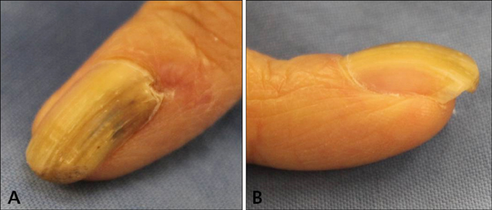
Fig. 1 (A) Yellowish, thickened nail plate of the right index finger, with splinter hemorrhages and longitudinal bands mimicking onychomycosis. (B) Lateral view of the nail plate showing longitudinal overcurvature.

Fig. 2 Ultrasonographic image showing a 2×5×7 mm hypoechogenic tumor under the proximal nail fold (A, B) and hyperechogenic villous projections of the nail matrix having low blood flow (C). (A) Transversal view. (B, C) Longitudinal view.

Fig. 3 (A) Intraoperative view of the filamentous tumor arising from the nail matrix. (B) Macroscopic appearance of the removed tumor with finger-like projections. (C) Proximal portion of the nail plate showing multiple cavities characteristic of onychomatricoma. (D) Normal nail plate growth 2 months after the excision.
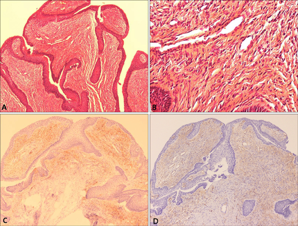
Fig. 4 (A, B) Histopathological section showing characteristic filiform projections lined by epithelium emerging from the nail matrix. (A) Medium-power view (H&E, ×100). (B) High-power view (H&E, ×400). (C) Immunohistochemical stain showing CD10 diffusely expressed in the stroma of the section. (D) Immunohistochemical stain showing CD34 diffusely expressed in the stroma of the section. (C, D) Medium-power view (×100).
Cited by 1 articles
-
Pigmented Onychomatricoma Showing a Longitudinal Melanonychia: A Case Report and Brief Review of Literature
Sung Cheol Jung, Tae Min Lee, Minsoo Kim, Gwanghyun Jo, Je-Ho Mun
Ann Dermatol. 2018;30(5):637-639. doi: 10.5021/ad.2018.30.5.637.
Reference
-
1. Baran R, Kint A. Onychomatrixoma. Filamentous tufted tumour in the matrix of a funnel-shaped nail: a new entity (report of three cases). Br J Dermatol. 1992; 126:510–515.
Article2. Tavares GT, Chiacchio NG, Chiacchio ND, Souza MV. Onychomatricoma: a tumor unknown to dermatologists. An Bras Dermatol. 2015; 90:265–267.
Article3. Di Chiacchio N, Tavares GT, Tosti A, Di Chiacchio NG, Di Santis E, Alvarenga L, et al. Onychomatricoma: epidemiological and clinical findings in a large series of 30 cases. Br J Dermatol. 2015; 173:1305–1307.
Article4. Perrin C, Baran R, Balaguer T, Chignon-Sicard B, Cannata GE, Petrella T, et al. Onychomatricoma: new clinical and histological features. A review of 19 tumors. Am J Dermatopathol. 2010; 32:1–8.
Article5. Gaertner EM, Gordon M, Reed T. Onychomatricoma: case report of an unusual subungual tumor with literature review. J Cutan Pathol. 2009; 36:Suppl 1. 66–69.
Article6. Tosti A, Piraccini BM, Calderoni O, Fanti PA, Cameli N, Varotti E. Onychomatricoma: report of three cases, including the first recognized in a colored man. Eur J Dermatol. 2000; 10:604–606.7. Baran R, Perrin C, Baudet J, Requena L. Clinical and histological patterns of dermatofibromas of the nail apparatus. Clin Exp Dermatol. 1994; 19:31–35.
Article8. Lee DY. The relation of onychomatricoma to onychodermis in the nail unit. Ann Dermatol. 2013; 25:394–395.
Article9. Piraccini BM, Antonucci A, Rech G, Starace M, Misciali C, Tosti A. Onychomatricoma: first description in a child. Pediatr Dermatol. 2007; 24:46–48.
Article10. Fayol J, Baran R, Perrin C, Labrousse F. Onychomatricoma with misleading features. Acta Derm Venereol. 2000; 80:370–372.
Article11. Cañueto J, Santos-Briz Á, García JL, Robledo C, Unamuno P. Onychomatricoma: genome-wide analyses of a rare nail matrix tumor. J Am Acad Dermatol. 2011; 64:573–578. 578.e1
Article12. Richert B, André J. Onychomatricoma. Ann Dermatol Venereol. 2011; 138:71–74.13. Miteva M, de Farias DC, Zaiac M, Romanelli P, Tosti A. Nail clipping diagnosis of onychomatricoma. Arch Dermatol. 2011; 147:1117–1118.
Article14. Soto R, Wortsman X, Corredoira Y. Onychomatricoma: clinical and sonographic findings. Arch Dermatol. 2009; 145:1461–1462.
Article15. Perrin C, Goettmann S, Baran R. Onychomatricoma: clinical and histopathologic findings in 12 cases. J Am Acad Dermatol. 1998; 39:560–564.
Article16. Hisaoka M, Hashimoto H. Elastofibroma: clonal fibrous proliferation with predominant CD34-positive cells. Virchows Arch. 2006; 448:195–199.
Article17. Misciali C, Iorizzo M, Fanti PA, Piraccini BM, Ceccarelli C, Santini D, et al. Onychoblastoma (hamartoma of the nail unit): a new entity? Br J Dermatol. 2005; 152:1077–1078.
Article18. Baran R. Is onychoblastoma really a new entity? Br J Dermatol. 2006; 154:384–385.
Article19. Yasuki Y. Acquired periungual fibrokeratoma--a proposal for classification of periungual fibrous lesions. J Dermatol. 1985; 12:349–356.20. Estrada-Chavez G, Vega-Memije ME, Toussaint-Caire S, Rangel L, Dominguez-Cherit J. Giant onychomatricoma: report of two cases with rare clinical presentation. Int J Dermatol. 2007; 46:634–636.
Article
- Full Text Links
-
- Actions
-
Cited
- CITED
-
- Close
- Share
-
- Similar articles
-
- CD13 Expression in Onychomatricoma: Association with Nail Matrix Onychodermis
- Three Cases of Nail Matrix Nevus in Children
- The Relation of Onychomatricoma to Onychodermis in the Nail Unit
- Thin Split-Thickness Toe Nail-Bed Grafts for Nail Bed Defects in Subungal Exostosis: Two Cases Report
- A Case of Multiple Periungual Fibrokeratoma with Matrix Differentiation
