Infect Chemother.
2012 Oct;44(5):382-385. 10.3947/ic.2012.44.5.382.
A Case of Pyogenic Liver Abscess Complicated by Hepatobronchial Fistula
- Affiliations
-
- 1Department of Internal Medicine, Soonchunhyang University College of Medicine, Cheonan, Korea. stevesh@sch.ac.kr
- KMID: 2170384
- DOI: http://doi.org/10.3947/ic.2012.44.5.382
Abstract
- Hepatobronchial fistula, an anatomic communication between the liver parenchyma and the bronchial tree, is a rare condition, which usually develops as a complication of amoebiasis, hydatid cysts, and trauma. We report on a case of a pyogenic liver abscess complicated by a hepatobronchial fistula, which responded well to treatment with antibiotics and percutaneous drainage. A 36-year-old male patient presented with a two-week history of dry cough, shortness of breath, right side abdominal pain, and fever. Chest computed tomography scan showed a heterogeneously enhanced abscess measuring approximately 6 cm in the right liver dome. Percutaneous drainage was performed and antibiotics were administered against Group C Streptococcus cultured from the abscess. After nine days of therapy, repositioning of the drainage catheter was performed and the patient coughed suddenly during injection of contrast media, and communication from abscess to bronchus was discovered. While maintaining abscess drainage and antibiotic therapy, the fistula diminished gradually and disappeared completely with resolution of the liver abscess.
Keyword
MeSH Terms
Figure
-

Figure 1 Chest radiograph shows costophrenic angle blunting at the right hemithorax with a small amount of fissural effusion and passive atelectasis in Rt. chest.
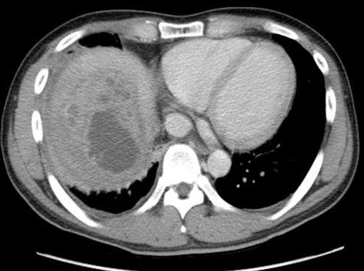
Figure 2 Chest computed tomography shows a huge heterogeneously enhanced mass with multiple septums measuring 5.9 cm×4.9 cm×3.5 cm at the right lobe of liver.
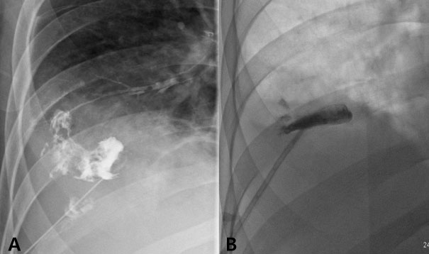
Figure 3 (A) Tubogram performed during repositioning of the pigtail catheter shows a fistula between the hepatic abscess and bronchial tree. (B) Follow-up tubogram taken after six weeks shows that the hepatobronchial fistula had disappeared.
Reference
-
1. Han SY. Liver abscess. Korean J Hepatol. 2006. 12(Supp 1):S5–S17.2. Borrie J, Shaw JH. Hepatobronchial fistula caused by hydatid disease. The Dunedin experience 1952-79. Thorax. 1981. 36:25–28.
Article3. Ibarra-Pérez C. Thoracic complications of amebic abscess of the liver: report of 501 cases. Chest. 1981. 79:672–677.
Article4. Tak WY, Jo CM, Keum MS, Kim DH, Kweon YO, Kim SK, Choi YH, Chung JM. A case of transcatheter arterial embolization-induced hepatobronchial fistula in a patient with hepatocellular carcinoma. Korean J Hepatol. 1999. 5:55–58.5. Sikma MA, Coenen JL, Kloosterziel C, Hasselt BA, Ruers TJ. A breakthrough in cryosurgery. Surg Endosc. 2002. 16:870.
Article6. Kim JH, Bae SH, Bae YS, Lee KJ, Hong JH, Yoon JB, Je IS. A hepatobronchial fistula in a patient with cholangiocarcinoma. Korean J Med. 2009. 77:Suppl 5. S1137–S1141.7. Jee MG, Choi YJ, Baik SK, Yoon SJ, Kim HS, Lee DK, Kim YJ. A case of hepatobronchial fistula in liver abscess. Korean J Med. 2003. 65:suppl 3. S703–S706.8. Okuda K, Kanda Y, Fukuyama Y, Sumikoshi T. Spontaneous hepatobronchial communications preceding pyothorax in a patient with suspected liver abscess. A case report. Gastroenterology. 1973. 65:124–129.
Article9. Moawad F, Truesdell A, Mulhall B. A "fishy" cough: hepatobronchial fistula due to a pyogenic liver abscess. N Z Med J. 2006. 119:U1906.10. Ala A, Safar-Aly H, Millar A. Metallic cough and pyogenic liver abscess. Eur J Gastroenterol Hepatol. 2001. 13:967–969.
Article11. Nesper M, McGahan JP. Hepatobronchial fistula with percutaneous pyogenic abscess drainage of the liver. Gastrointest Radiol. 1985. 10:129–131.
Article12. Huch Böni RA, Peter J, Marincek B. Amebic abscess of the liver manifested by "hemoptysis": US, CT, and MRI findings. Abdom Imaging. 1995. 20:214–216.
Article
- Full Text Links
-
- Actions
-
Cited
- CITED
-
- Close
- Share
-
- Similar articles
-
- A case of hepatobronchial fistula in liver abscess
- Hepatobronchial Fistula and Lung Abscess after Transarterial Chemoembolization
- A Case of Transcatheter Arterial Embolization-nduced Hepatobronchial Fistula in a Patient with Hepatocellular Carcinoma
- A Case of Pyogenic Liver Abscess Complicated with Endophthalmitis
- Pyogenic Liver Abscess by Proteus vulgaris Complicated with Acute Purulent Pericarditis: A Case Report and Review of Korean Literature
