Endocrinol Metab.
2011 Sep;26(3):272-275. 10.3803/EnM.2011.26.3.272.
A Case of Adrenal Teratoma
- Affiliations
-
- 1Department of Internal Medicine, Division of Endocrinology and Metabolism, Konyang University Hospital, Daejeon, Korea.
- 2Department of Internal Medicine, Division of Endocrinology and Metabolism, Konyang University College of Medicine, Daejeon, Korea. kbjoon4u@hananet.net
- KMID: 2169182
- DOI: http://doi.org/10.3803/EnM.2011.26.3.272
Abstract
- Teratoma is a congenital tumor containing tissues derived from all germ layers. Teratoma in the region of the adrenal gland is a very uncommon retroperitoneal tumor. Only 7 cases of adrenal teratoma have been reported worldwide, but in Korea, no similar cases have been reported until now. This case report describes an adrenal teratoma in a 38-year-old healthy woman who was incidentally diagnosed with a left adrenal mass on abdominal ultrasonography during a medical inspection. Computed tomographic scans revealed a 9-cm heterogeneous circumscribed round mass, containing primarily fat tissue, and a solid calcification component in the left adrenal gland. Adrenal hormonal assessment results and biochemical markers for gonadal neoplasia were negative. Result of serum laboratory tests were normal. The patient underwent laparoscopic adrenalectomy. Histologic analysis confirmed the diagnosis of a mature teratoma; the obtained specimen measured 5 x 7 x 7.5 cm and weighed 267 g. The surface of the mass was smooth, and sebaceous tissue and hair with hard material were observed on the incisional surface. The patient was discharged on postoperative day 4, without complications. In this case report, we describe the incidental finding of a teratoma occurring in the adrenal gland region in a healthy woman; the teratoma was laparoscopically excised.
MeSH Terms
Figure
-
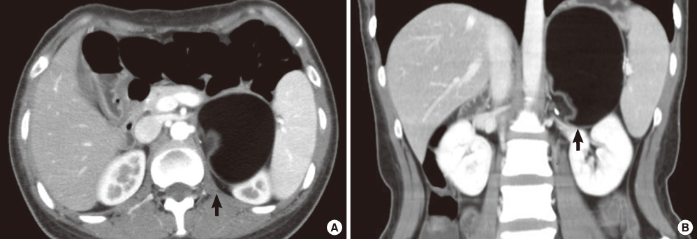
Fig. 1 CT finding of adrenal teratoma. A. Cross section. B. Sagittal section. Two abdominal CT shows a circumscribed round mass, containing with mainly fat tissue, and calcification, solid component in left adrenal gland (arrow).
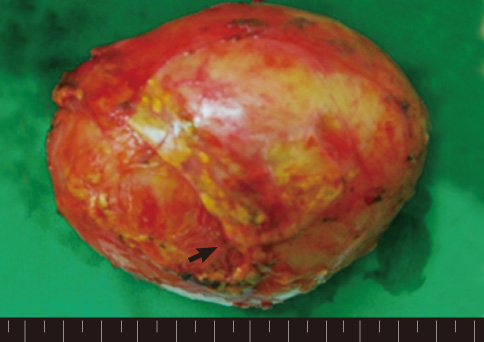
Fig. 2 Gross findings of the excised adrenal mass. Specimen measured 5 × 7 × 7.5 cm and weighed 267 g. Remnant portion of adrenal gland were shown (arrow). The surface of the mass was smooth, and sebaceous tissue and hair with hard material were seen on the incisional surface.

Fig. 3 Histology confirms the diagnosis of benign mature teratoma (H&E, × 100). A. Muscle (arrow). B. Skin and skin appendage, neural tissue, hair follicle (arrow). C. Mucous cyst (arrow).
Reference
-
1. Hui JPK, Luk WH, Siu CW, Chan JCS. Teratoma in the region of an adrenal gland in a 77-year-old man. J HK Coll Radiol. 2004. 7:206–209.2. Bruneton JN, Diard F, Drouillard JP, Sabatier JC, Tavernier JF. Primary retroperitoneal teratoma in adults: presentation of two cases and review of the literature. Radiology. 1980. 134:613–616.3. Polo JL, Villarejo PJ, Molina M, Yuste P, Menendez JM, Babe J, Puente S. Giant mature cystic teratoma of the adrenal region. AJR Am J Roentgenol. 2004. 183:837–838.4. Lam KY, Lo CY. Teratoma in the region of adrenal gland: a unique entity masquerading as lipomatous adrenal tumor. Surgery. 1999. 126:90–94.5. Castillo OA, Vitagliano G, Villeta M, Arellano L, Santis O. Laparoscopic resection of adrenal teratoma. JSLS. 2006. 10:522–524.6. Parnes LS, Sun AH. Teratoma of the middle ear. J Otolaryngol. 1995. 24:165–167.7. Gonzalez-Crussi F. Gonzalez-Crussi F, editor. Nomenclature. Extragonal teratomas, fasc 18, ser 2. 1982. Washington DC: Armed Forces Institute of Pathology;1–9.8. Saiga T, Osasa H, Hatayama H, Miyamoto T, Ono H, Mikami T. The origin of extragonadal teratoma: case report of an immature teratoma occurring in a prenatal brain. Pediatr Pathol. 1991. 11:759–770.9. Gatcombe HG, Assikis V, Kooby D, Johnstone PA. Primary retroperitoneal teratomas: a review of the literature. J Surg Oncol. 2004. 86:107–113.10. Tezel E, Sare M, Edali N, Oguz M, Uluoglu O, Gokok NH. Retroperitoneal malignant teratoma. A case report. Mater Med Pol. 1995. 27:123–125.11. Goyal M, Sharma R, Sawhney P, Sharma MC, Berry M. The unusual imaging appearance of primary retroperitoneal teratoma: report of a case. Surg Today. 1997. 27:282–284.12. Rais-Bahrami S, Varkarakis IM, Lujan G, Jarrett TW. Primary retroperitoneal teratoma presenting as an adrenal tumor in an adult. Urology. 2007. 69:185.e1–185.e2.13. Davidson AJ, Hartman DS, Goldman SM. Mature teratoma of the retroperitoneum: radiologic, pathologic, and clinical correlation. Radiology. 1989. 172:421–425.14. Lam KY, Lo CY. Adrenal lipomatous tumours: a 30 year clinicopathological experience at a single institution. J Clin Pathol. 2001. 54:707–712.
