Distribution of CD10-positive epithelial and mesenchymal cells in human mid-term fetuses: a comparison with CD34 expression
- Affiliations
-
- 1Department of Anatomy, Chonbuk National University Medical School, Jeonju, Korea.
- 2Department of Surgery, Daejeon Sun Hospital, Daejeon, Korea.
- 3Department of Surgery and Biomedical Research Institute, Chonbuk National University Hospital, Chonbuk National University Medical School, Jeonju, Korea. chobh@jbnu.ac.kr
- 4Division of Otorhinolaryngology, Sendai Municipal Hospital, Sendai, Japan.
- 5Division of Internal Medicine, Iwamizawa Kojin-kai Hospital, Iwamizawa, Japan.
- KMID: 2168854
- DOI: http://doi.org/10.5115/acb.2014.47.1.28
Abstract
- CD10, a marker of immature B lymphocytes, is expressed in the developing epithelium of mammary glands, hair follicles, and renal tubules of human fetuses. To assess mesenchymal and stromal expression of CD10, we performed immunohistochemical assays in whole body sections from eight fetuses of gestational ages 15-20 weeks. In addition to expression in urinary tract and intestinal epithelium, CD10 was strongly expressed at both gestational ages in fibrous tissues surrounding the airways from the larynx to lung alveoli, in the periosteum and ossification center, and in the glans of external genitalia. CD10 was not expressed, however, in other cavernous tissues. These findings suggest that mesenchymal, in addition to epithelial cells at specific sites, are likely to express CD10. The glomeruli, alveoli, and glans are all end products of budding or outgrowth processes in the epithelium or skin. However, in contrast to the CD34 marker of stromal stem cells, CD10 was not expressed in vascular progenitor cells and in differentiated vascular endothelium. The alternating pattern of CD10 and CD34 expression suggests that these factors play different roles in cellular differentiation and proliferation of the kidneys, airway and external genitalia.
Keyword
MeSH Terms
Figure
-
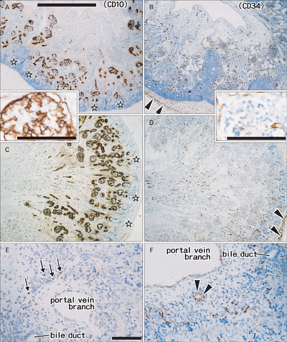
Fig. 1 Immunohistochemistry of CD10 (A, C, E) and CD34 (B, D, F) in the kidney and liver. Panels (A) and (B) (near sections) show a kidney of gestational age (GA) 15 weeks, whereas panels (C) and (D) (near sections) show kidneys at GA 20 weeks. Inserts between panels (A) and (C) and between panels (B) and (D) (scale bars=0.1 mm) show more highly magnified views of a glomerulus in panels (C) and (D). Panels (E) and (F) (near sections) show the liver parenchyma around a portal vein branch at 15 weeks. The superficial layer of the renal cortex (stars in panels A and C) was negative for CD10. Renal capsules were positive for CD34 (arrowheads in panels B and D). CD10-positive immature B lymphocytes were present in a portal vein branch (arrows in panel E), whereas CD34-positive cells formed rosette-like structures (arrows in panel F). Panels (A-D) (E and F) were prepared at the same magnification. Scale bars in panels (A) and (E)=1 mm (A-D), 0.1 mm (E, F).

Fig. 2 Immunohistochemical expression of CD10 (A, C, E) and CD34 (B, D, F) in the intestines, pancreas, and stomach. Panels (A) and (B) (near sections) show the ileum at gestational age (GA) 15 weeks, whereas panels (C) and (D) (near sections) show the duodenum and pancreas at GA 20 weeks. Panels (E) and (F) (near sections) show the stomach at GA 15 weeks. CD10 was positive at the luminal surface of the intestinal villi (A, C), but not in the stomach (E). CD10-positive immature B lymphocytes are shown in panel (C). (B, D) The mesentery and pancreas showed strong expression of CD34. SMA, superior mesenteric artery. All panels were prepared at the same magnification. Scale bar in panel (A)=1 mm (A-F).
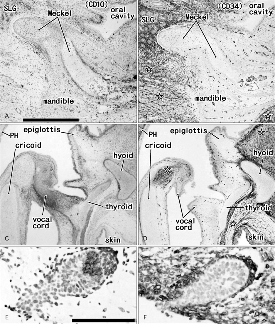
Fig. 3 Immunohistochemical expression of CD10 (A, C, E) and CD34 (B, D, F) in the mandible, larynx, and hair. Panels (A) and (B) (near sections) show the mandible containing Meckel's cartilage from a fetus of gestational age (GA) 15 weeks, and panels (C) and (D) (near sections) show the larynx of the same fetus. Panels (E) and (F) (near sections) show a hair follicle in the anterior thoracic skin at GA 20 weeks. CD10-positive fibrous tissues were present in the larynx (C) and along Meckel's cartilage (A), whereas CD34-positive fibrous tissues were abundant in striated muscles (stars in panels B and D), the sublingual gland (SLG in panel B) and subcutaneous tissue (D, F). PH, epithelium of the posterior wall of the pharynx. Panels (A-D) (or E and F) were prepared at the same magnification. Scale bars in panels (A) and (E)=1 mm (A-D), 0.1 mm (E, F).
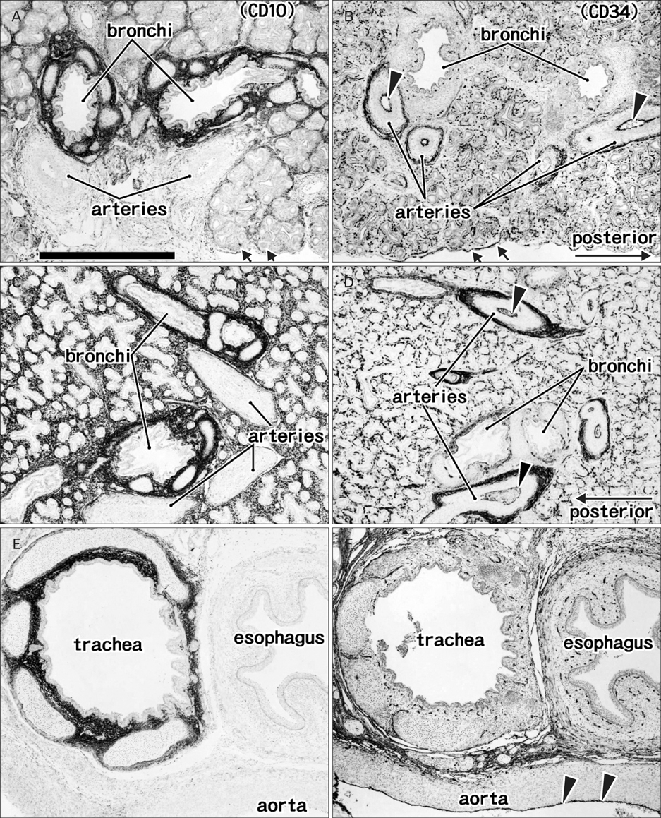
Fig. 4 Immunohistochemical expression of CD10 (A, C, E) and CD34 (B, D, F) in the lungs and mediastinum. Panels (A) and (B) (near sections) show the right pulmonary hilus at gestational age (GA) 15 weeks, while panels (C) and (D) (near sections) show branching of the subsegmental bronchi and arteries in segment VI of the right lung at GA 20 weeks. The bronchi are surrounded by CD10-positive fibrous tissues, whereas the arteries are surrounded by CD34-positive tissues. The connective tissues of the trachea also express CD10 strongly. The endothelium of the aorta and pulmonary arteries express CD34 (arrowheads in panels B, D, and F). Panels (E) and (F) (near sections) show mediastinal structures at GA 15 weeks. All panels were prepared at the same magnification. Scale bar in panel (A)=1 mm (A-F).
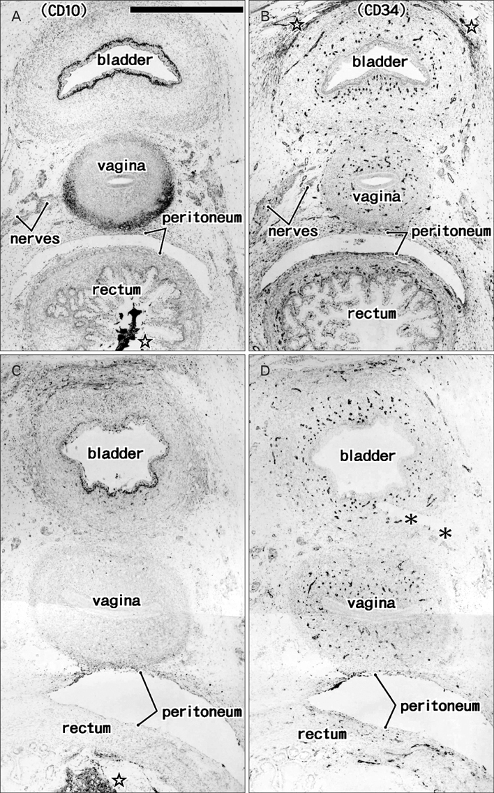
Fig. 5 Immunohistochemical expression of CD10 (A, C) and CD34 (B, D) in the bladder, vagina, and rectum. Panels (A) and (B) (near sections) show the bladder, vagina, and rectum at gestational age (GA) 15 weeks, while panels (C) and (D) (adjacent sections) show these three structures at GA 20 weeks. CD10 expression was observed in the bladder epithelium and the adventitia of the vagina, whereas the thin vessels and retropubic fibrous tissue were positive for CD34 (stars in panel B). A CD10-positive meconium-like substance was observed in the rectum (stars in panels A and C). Asterisks in panel (D) indicate an artifactual cleft generated during the histological procedure. All panels were prepared at the same magnification. Scale bar in panel (A)=1 mm (A-D).
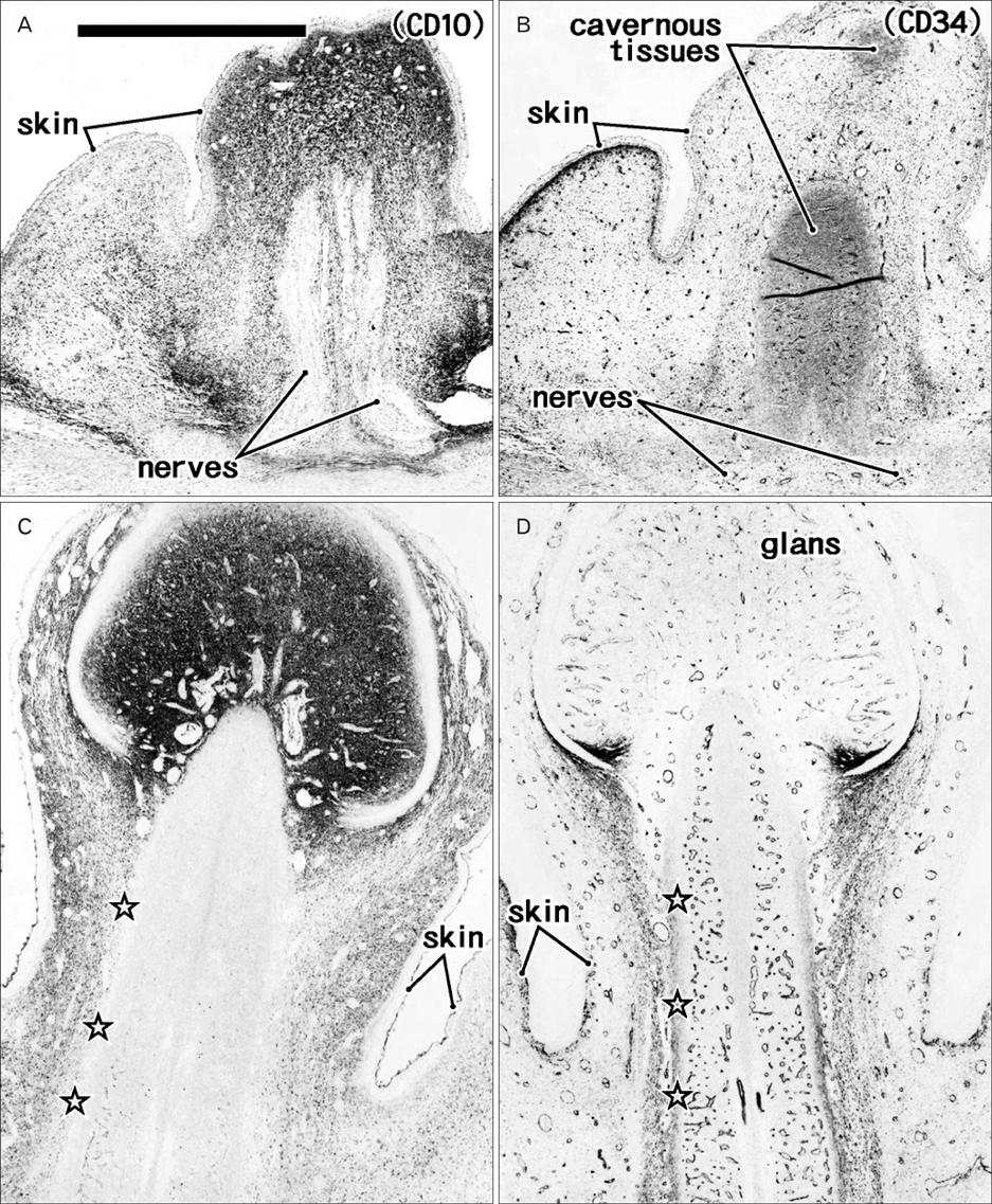
Fig. 6 Immunohistochemical expression of CD10 (A, C) and CD34 (B, D) in the clitoris. Panels (A) and (B) (near sections) display the clitoris and labium at gestational age (GA) 15 weeks, while panels (C) and (D) (adjacent sections) show the clitoris at GA 20 weeks. CD10-positive fibrous tissues were present in the subcutaneous tissue and glans of the clitoris, whereas CD34 was positive in thin vessels and in some CD10-positive fibrous tissues. The tunica albuginea of cavernous tissues was positive for CD34 (stars in panel D), but negative for CD10 (stars in panel C). All panels were prepared at the same magnification. Scale bar in panel (A)=1 mm (A-D).

Fig. 7 Immunohistochemical expression of CD10 (A, C, E) and CD34 (B, D, F) in bones, joints and muscles. Panels (A) and (B) (near sections) show the ilium at gestational age (GA) 15 weeks, panels (C) and (D) (adjacent sections) show the hip joint at GA 20 weeks, and panels (E) and (F) (adjacent sections) show the gluteal muscles and the skin covering them at GA 20 weeks. The endochondral ossification center (stars in panels A and B) as well as the synovial tissue (panels C and D) were positive for both CD10 and CD34. Striated muscle fibers were separated by abundant sheaths strongly positive for CD34 (F) and weakly positive for CD10 (E). All panels were prepared at the same magnification. Scale bar in panel (A)=1 mm (A-F).
Reference
-
1. Bárcena A, Muench MO, Galy AH, Cupp J, Roncarolo MG, Phillips JH, Spits H. Phenotypic and functional analysis of T-cell precursors in the human fetal liver and thymus: CD7 expression in the early stages of T- and myeloid-cell development. Blood. 1993; 82:3401–3414.2. Paramithiotis E, Cooper MD. Memory B lymphocytes migrate to bone marrow in humans. Proc Natl Acad Sci U S A. 1997; 94:208–212.3. D'Arena G, Musto P, Cascavilla N, Di Giorgio G, Fusilli S, Zendoli F, Carotenuto M. Flow cytometric characterization of human umbilical cord blood lymphocytes: immunophenotypic features. Haematologica. 1998; 83:197–203.4. Poblet E, Jiménez F. CD10 and CD34 in fetal and adult human hair follicles: dynamic changes in their immunohistochemical expression during embryogenesis and hair cycling. Br J Dermatol. 2008; 159:646–652.5. Bachelard-Cascales E, Chapellier M, Delay E, Pochon G, Voeltzel T, Puisieux A, Caron de Fromentel C, Maguer-Satta V. The CD10 enzyme is a key player to identify and regulate human mammary stem cells. Stem Cells. 2010; 28:1081–1088.6. Faa G, Gerosa C, Fanni D, Nemolato S, Marinelli V, Locci A, Senes G, Mais V, Van Eyken P, Iacovidou N, Monga G, Fanos V. CD10 in the developing human kidney: immunoreactivity and possible role in renal embryogenesis. J Matern Fetal Neonatal Med. 2012; 25:904–911.7. Waller EK, Huang S, Terstappen L. Changes in the growth properties of CD34+, CD38- bone marrow progenitors during human fetal development. Blood. 1995; 86:710–718.8. Abe S, Suzuki M, Cho KH, Murakami G, Cho BH, Ide Y. CD34-positive developing vessels and other structures in human fetuses: an immunohistochemical study. Surg Radiol Anat. 2011; 33:919–927.9. Katori Y, Kiyokawa H, Kawase T, Murakami G, Cho BH. CD34-positive primitive vessels and other structures in human fetuses: an immunohistochemical study. Acta Otolaryngol. 2011; 131:1086–1090.10. Chang H, Cho KH, Hayashi S, Kim JH, Abe H, Rodriguez-Vazquez JF, Murakami G. Site- and stage-dependent differences in vascular density of the human fetal brain. Childs Nerv Syst. 2014; 30:399–409.11. Yajin S, Murakami G, Takeuchi H, Hasegawa T, Kitano H. The normal configuration and interindividual differences in intramural lymphatic vessels of the esophagus. J Thorac Cardiovasc Surg. 2009; 137:1406–1414.12. Moon WS, Cho BH, Hayashi S, Kim JH, Murakami G, Fukuzawa Y, Nakano T. Cytokeratin-positive hepatocytes in the hilar region: an immunohistochemical study using livers from fetuses and elderly individuals. Ann Anat. 2011; 193:224–230.13. Gerlach JC, Over P, Turner ME, Thompson RL, Foka HG, Chen WC, Péault B, Gridelli B, Schmelzer E. Perivascular mesenchymal progenitors in human fetal and adult liver. Stem Cells Dev. 2012; 21:3258–3269.14. Kano Y, Sakurai H, Shidara J, Toida S, Yasuda H. Histopathological and immunohistochemical studies of acquired tracheobronchomalacia: an autopsy case report. ORL J Otorhinolaryngol Relat Spec. 1996; 58:288–294.15. Carden KA, Boiselle PM, Waltz DA, Ernst A. Tracheomalacia and tracheobronchomalacia in children and adults: an in-depth review. Chest. 2005; 127:984–1005.16. Invernici G, Ponti D, Corsini E, Cristini S, Frigerio S, Colombo A, Parati E, Alessandri G. Human microvascular endothelial cells from different fetal organs demonstrate organ-specific CAM expression. Exp Cell Res. 2005; 308:273–282.
- Full Text Links
-
- Actions
-
Cited
- CITED
-
- Close
- Share
-
- Similar articles
-
- Characterization of mesenchymal cells beneath cornification of the fetal epithelium and epidermis at the face: an immunohistochemical study using human fetal specimens
- Comparative Evaluation for Potential Differentiation of Endothelial Progenitor Cells and Mesenchymal Stem Cells into Endothelial-Like Cells
- Effects of Damaged Human Corneal Epithelial Cells on Differentiation of Human Mesenchymal Stem Cell
- Immunohistochemical Phenotypes of Phyllodes Tumor of the Breast
- A temporary disc-like structure at the median atlanto-axial joint in human fetuses
